Japanese
English
- 有料閲覧
- Abstract 文献概要
- 1ページ目 Look Inside
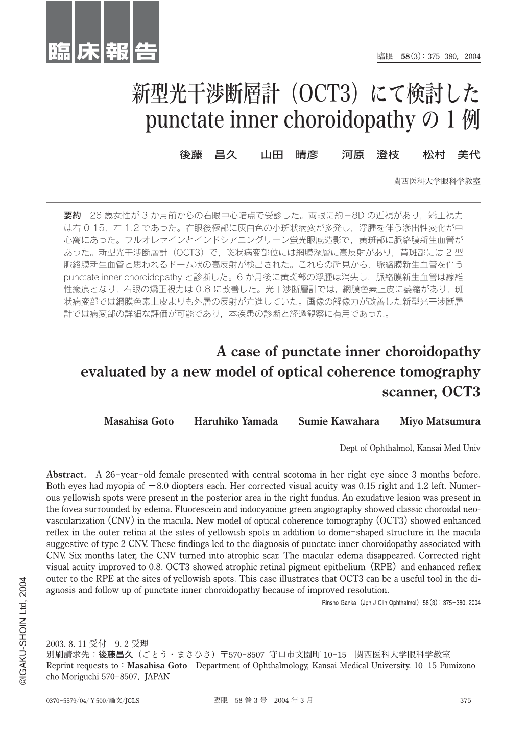
26歳女性が3か月前からの右眼中心暗点で受診した。両眼に約-8Dの近視があり,矯正視力は右0.15,左1.2であった。右眼後極部に灰白色の小斑状病変が多発し,浮腫を伴う滲出性変化が中心窩にあった。フルオレセインとインドシアニングリーン蛍光眼底造影で,黄斑部に脈絡膜新生血管があった。新型光干渉断層計(OCT3)で,斑状病変部位には網膜深層に高反射があり,黄斑部には2型脈絡膜新生血管と思われるドーム状の高反射が検出された。これらの所見から,脈絡膜新生血管を伴うpunctate inner choroidopathyと診断した。6か月後に黄斑部の浮腫は消失し,脈絡膜新生血管は線維性瘢痕となり,右眼の矯正視力は0.8に改善した。光干渉断層計では,網膜色素上皮に萎縮があり,斑状病変部では網膜色素上皮よりも外層の反射が亢進していた。画像の解像力が改善した新型光干渉断層計では病変部の詳細な評価が可能であり,本疾患の診断と経過観察に有用であった。
A 26-year-old female presented with central scotoma in her right eye since 3months before. Both eyes had myopia of-8.0 diopters each. Her corrected visual acuity was 0.15 right and 1.2 left. Numerous yellowish spots were present in the posterior area in the right fundus. An exudative lesion was present in the fovea surrounded by edema. Fluorescein and indocyanine green angiography showed classic choroidal neovascularization(CNV)in the macula. New model of optical coherence tomography(OCT3)showed enhanced reflex in the outer retina at the sites of yellowish spots in addition to dome-shaped structure in the macula suggestive of type 2 CNV. These findings led to the diagnosis of punctate inner choroidopathy associated with CNV. Six months later,the CNV turned into atrophic scar. The macular edema disappeared. Corrected right visual acuity improved to 0.8. OCT3 showed atrophic retinal pigment epithelium(RPE)and enhanced reflex outer to the RPE at the sites of yellowish spots. This case illustrates that OCT3 can be a useful tool in the diagnosis and follow up of punctate inner choroidopathy because of improved resolution.

Copyright © 2004, Igaku-Shoin Ltd. All rights reserved.


