Japanese
English
- 有料閲覧
- Abstract 文献概要
- 1ページ目 Look Inside
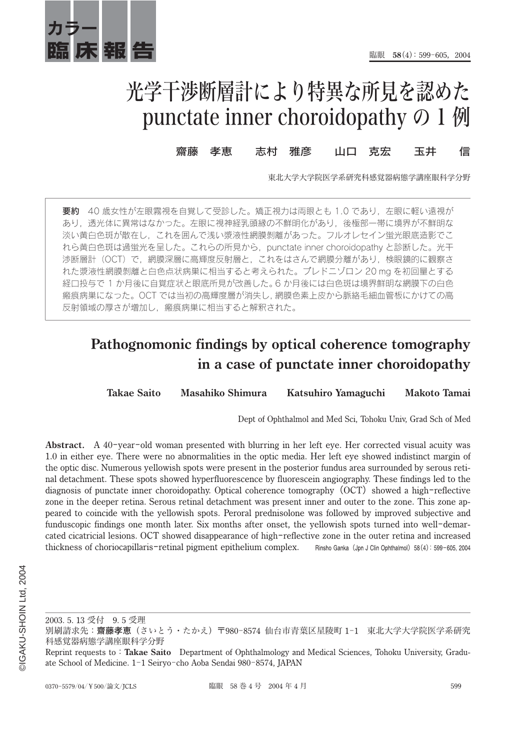
40歳女性が左眼霧視を自覚して受診した。矯正視力は両眼とも1.0であり,左眼に軽い遠視があり,透光体に異常はなかった。左眼に視神経乳頭縁の不鮮明化があり,後極部一帯に境界が不鮮明な淡い黄白色斑が散在し,これを囲んで浅い漿液性網膜剝離があった。フルオレセイン蛍光眼底造影でこれら黄白色斑は過蛍光を呈した。これらの所見から,punctate inner choroidopathyと診断した。光干渉断層計(OCT)で,網膜深層に高輝度反射層と,これをはさんで網膜分離があり,検眼鏡的に観察された漿液性網膜剝離と白色点状病巣に相当すると考えられた。プレドニゾロン20mgを初回量とする経口投与で1か月後に自覚症状と眼底所見が改善した。6か月後には白色斑は境界鮮明な網膜下の白色瘢痕病巣になった。OCTでは当初の高輝度層が消失し,網膜色素上皮から脈絡毛細血管板にかけての高反射領域の厚さが増加し,瘢痕病巣に相当すると解釈された。
A 40-year-old woman presented with blurring in her left eye. Her corrected visual acuity was 1.0 in either eye. There were no abnormalities in the optic media. Her left eye showed indistinct margin of the optic disc. Numerous yellowish spots were present in the posterior fundus area surrounded by serous retinal detachment. These spots showed hyperfluorescence by fluorescein angiography. These findings led to the diagnosis of punctate inner choroidopathy. Optical coherence tomography(OCT)showed a high-reflective zone in the deeper retina. Serous retinal detachment was present inner and outer to the zone. This zone appeared to coincide with the yellowish spots. Peroral prednisolone was followed by improved subjective and funduscopic findings one month later. Six months after onset,the yellowish spots turned into well-demarcated cicatricial lesions. OCT showed disappearance of high-reflective zone in the outer retina and increased thickness of choriocapillaris-retinal pigment epithelium complex.
Copyright © 2004, Igaku-Shoin Ltd. All rights reserved.