Japanese
English
- 有料閲覧
PPV購入案内
1,320円 (1,200円+税10%) こちらは、1251ページから1255ページの文献です。購入には医書.jp本会員登録が必要です。
会員登録完了後に改めて上記「購入サイトへ」を選択してください。
または「購入サイトへ」選択後に「購入ログイン」、「新規会員登録」の順に進んでください。
会員登録について詳しくはをご確認ください。
- Abstract 文献概要
- 1ページ目 Look Inside
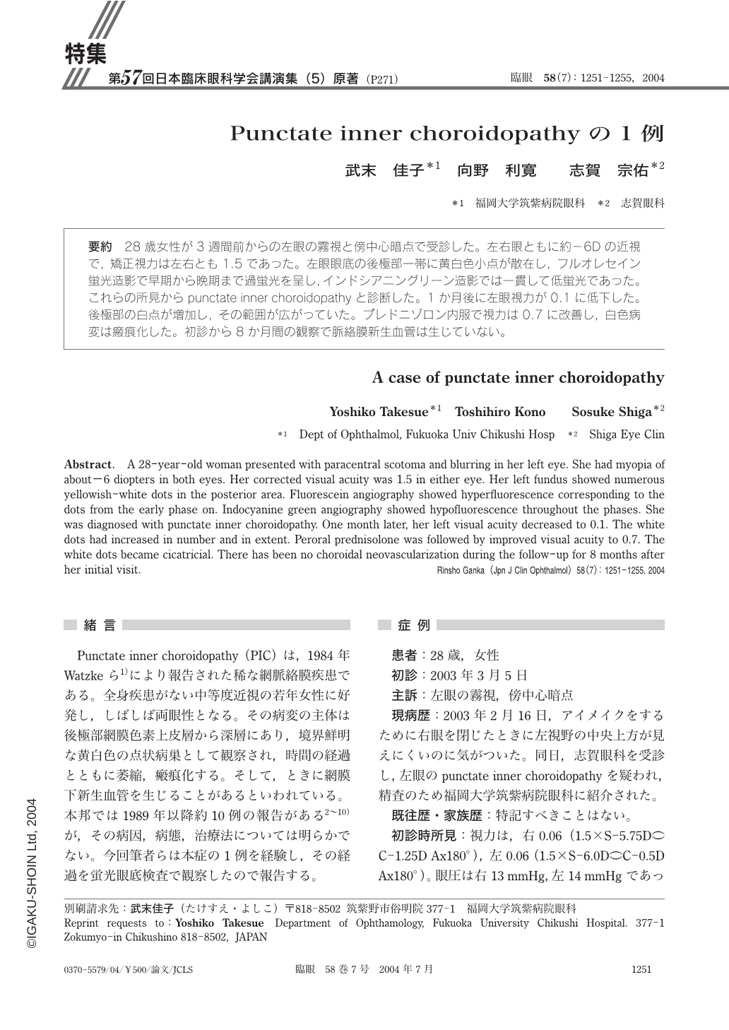
28歳女性が3週間前からの左眼の霧視と傍中心暗点で受診した。左右眼ともに約-6Dの近視で,矯正視力は左右とも1.5であった。左眼眼底の後極部一帯に黄白色小点が散在し,フルオレセイン蛍光造影で早期から晩期まで過蛍光を呈し,インドシアニングリーン造影では一貫して低蛍光であった。これらの所見からpunctate inner choroidopathyと診断した。1か月後に左眼視力が0.1に低下した。後極部の白点が増加し,その範囲が広がっていた。プレドニゾロン内服で視力は0.7に改善し,白色病変は瘢痕化した。初診から8か月間の観察で脈絡膜新生血管は生じていない。
A 28-year-old woman presented with paracentral scotoma and blurring in her left eye. She had myopia of about-6 diopters in both eyes. Her corrected visual acuity was 1.5 in either eye. Her left fundus showed numerous yellowish-white dots in the posterior area. Fluorescein angiography showed hyperfluorescence corresponding to the dots from the early phase on. Indocyanine green angiography showed hypofluorescence throughout the phases. She was diagnosed with punctate inner choroidopathy. One month later,her left visual acuity decreased to 0.1. The white dots had increased in number and in extent. Peroral prednisolone was followed by improved visual acuity to 0.7. The white dots became cicatricial. There has been no choroidal neovascularization during the follow-up for 8months after her initial visit.
Copyright © 2004, Igaku-Shoin Ltd. All rights reserved.