Japanese
English
- 有料閲覧
PPV購入案内
1,320円 (1,200円+税10%) こちらは、311ページから319ページの文献です。購入には医書.jp本会員登録が必要です。
会員登録完了後に改めて上記「購入サイトへ」を選択してください。
または「購入サイトへ」選択後に「購入ログイン」、「新規会員登録」の順に進んでください。
会員登録について詳しくはをご確認ください。
- Abstract 文献概要
- 1ページ目 Look Inside
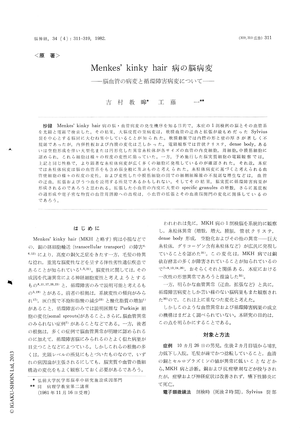
抄録 Menkes’kinky hair病の脳・血管病変の発生機序を知る目的で,本症の1剖検例の脳とその血管系を光顕と電顕で検索した。その結果,大脳皮質の巣病変は,軟膜血管の迂曲と拡張が最もめだったSylvius裂を中心とする脳回に大むね集中していることが知られた。軟膜動脈では内腔の形と壁の厚さが著しく不規則であったが,内弾性板および内膜の変化は乏しかった。電顕観察では管状クリステ,dense body,あるいは空胞形成を伴い大型化または円形化した異常糸粒体が各サイズの血管の内皮細胞,周細胞,中膜筋細胞に認められ,これら細胞は様々の程度の変性に陥っていた。一方,予め施行した脳実質細胞の電顕観察では,上記と同じ性格で,より顕著な糸粒体病変が広く多くの細胞に発現しているのが確認された。それ故,本症では糸粒体病変は脳の血管系をも含め脳全般に及ぶものと考えられた。糸粒体病変に基づくと考えられる血管壁細胞の様々の程度の変性,および変性した中膜筋細胞の間での細網細線維の不規則な増生などは,血管の迂曲,拡張およびうつ血を説明する所見であるかもしれない。そしてその結課,脳実質に循環障害病巣が形成されるのであろうと思われる。拡張した小血管の内皮に大型のspecific granulesの増数,さらに基底板の過形成や電子密な物質の血管周囲隙への出現は,小血管の拡張とその血液脳関門の変化に関係しているのであろう。
In order to elucidate the mechanism of develop-ment of cerebrovascular lesions in Menkes' kinky hair disease (MKHD), the authors examined the brain and its vasculature of an autopsied case of this disease in light and electron microscopy. The gross and light microscopic examinations revealed that focal degeneration including pseudolaminar necrosis of cerebral cortices roughly concentrated on the gyri in and around the Sylvian fissures where tortuous vessels with congestion and dilata-tion were most prominently present.
Despite their conspicuous irregularity in shape of the lumens and in thickness of the walls, the vessels were devoid of significant changes in elastic lamina and intima.
In contrast, mitochondrial disease manifested by enlargement and/or rounding of mitochondria with tubular cristae and dense body as well as vacuolar degeneration were noted in various sizes of vessels in endothelial cells, pericytes and medial muscle cells, which exibited various degrees of degenera-tion. On the other hand the preliminary observa-tion had disclosed the mitochondrial changes of the same character in the neurons in various structures of this brain, such as Purkinje cells, granule cells, and neurons in cerebral cortex, thalamus and globus pallidus, and even in glial cells. Therefore it seemed reasonable to consider that the mitochon-drial disease in MKHD might be ubiquituous in the brain including its vasculature.
The observations of various degrees of degenera-tion of vessel wall cells due to mitochondrial disease and of irregular proliferation of reticulin fibrils in the spaces among degenerated muscle cells of the tunica media may be the evidences responsible for the tortuosity, dilatation and congestion of vessels, which eventually give rise to the vascular lesions in the brain parenchyme in MKHD.
The findings of increased numbers of enlarged specific granules in the endothelium of dilated ves-sels, and dense material accumulation in their perivascular spaces with proliferation of basal lamina may have something to do with dilatation of small vessels and alteration of their blood brain barrier.
Copyright © 1982, Igaku-Shoin Ltd. All rights reserved.