Japanese
English
- 有料閲覧
- Abstract 文献概要
- 1ページ目 Look Inside
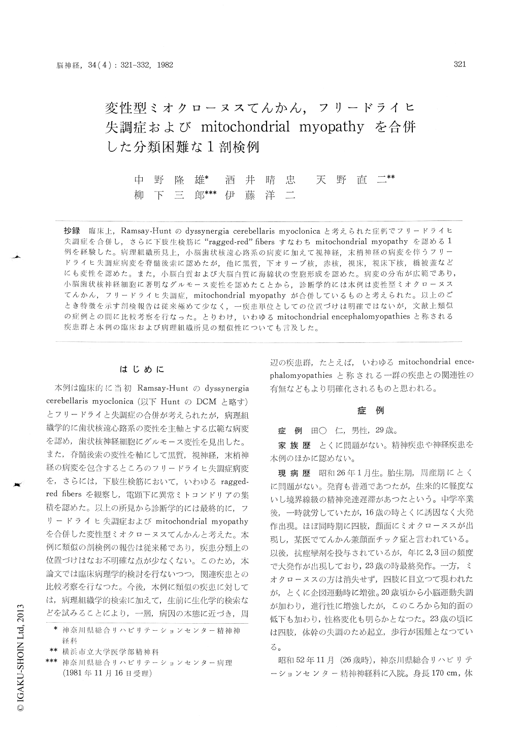
抄録 臨床上,Ramsay-Huntのdyssynergia cerebellaris rnyoclonicaと考えられた症例でブリードライヒ失調症を合併し,さらに下肢生検筋に"ragged-red"fibersすなわちmitochondrial myopathyを認める1例を経験した。病理組織所見上,小脳歯状核遠心路系の病変に加えて視神経,末梢神経の病変を伴うフリードライヒ失調症病変を脊髄後索に認めたが,他に黒質,下オリーブ核,赤核,視床,視床下核,橋被蓋などにも変性を認めた、また,小脳白質および大脳白質に海綿状の空胞形成を認めた。病変の分布が広範であり,小脳歯状核神経細胞に著明なグルモース変性を認めたことから,診断学的には本例は変性型ミオクローヌスてんかん,フリードライヒ失調症,mitochondrial myopathyが合併しているものと考えられた。以上のごとき特徴を示す剖検報告は従来極めて少なく,一疾患単位としての位置づけは明確ではないが,文献上類似の症例との間に比較考察を行なった。とりわけ,いわゆるmitochondrial encephalomyopathiesと称される疾患群と本例の臨床および病理組織所見の類似性についても言及した。
The patient was born in 1951. Mild mental re-tardation became manifested in infancy. There were no hereditary and familial histories. Since age of 16 years, generalized convulsions and myoclonic jerks emerged and resistant to anticonvulsive therapy. Since age of 20 years, cerebellar ataxia developed and mental deterioration became stand out. At the age of 23, he could not stand up without support due to ataxia and admitted to the hospital at the age of 26.
Adding to cerebellar ataxia and intentional myo-clonus, marked hypotonia of the skeletal muscles and remarkable diminutions of both deep tendon reflexes and deep sensations predominating in the lower extremities were revealed neurologically. Noticeable muscle atrophy was observed in the distal part of the lower extremities and to the lesser extent in the upper extremities. The feet showed pes cavus. At this point, Ramsay-Hunt syndrome combined with Friedreich's ataxia was suspected.
Afterward, decrease visual acuity and optic nerve atrophy were noticed by ophthalmologic examina-tion. However, disturbance of ocular movement or ptosis could not be detected. At the age of 28, atrophic muscle of the leg and sural nerve were biopsied. The biopsied muscles were histochemically studied and demonstrated to have many "ragged-red" fibers stained positively with Gomori trichrome. Electron microscopic study of these "ragged-red" fibers revealed unusual accumulation of abnormal mitochodria beneath the sarcolemma.
Abnormal mitochodrias were divided into two main types : The one type mitochondrias included peculior paracrystalline inclusions in their matrix and the others showed whirl-like cristae. These characteristic features of abnormal mitochondrias were virtually identical to those of Kearns-Shy syndrome or ophthalmoplegia plus and so on. Biopsied sural nerve was markedly demyelinated with distinct axonal degeneration.
Laboratory examinations revealed moderately elevated CSF protein, serum GOT, GPT, LDH and CPK. Serum CPK elevated temporarily to 2, 376 I. U.. Electron encephalogram showed severe epi-leptic pattern.
Post-mortem findings: The brain weighed 1, 140g.The cerebellum, brain stem and spinal cord were mildly atrophic. Ventricular systems showed diffuse and moderate dilatation, especially marked at the fourth ventricle. Dentate nucleus was severely shrunken and discolored brownish. Substantia nigra was moderately depigmentated. The tegmentum of pons was noticed to be unusually narrowed. Neurons of the dentate nucleus were severely lost with glial proliferation. Marked grumose degenera-tion was noted. Noticeable isomorphic fibrous gliosis was seen in the hilus of dentate nucleus, superior cerebellar peduncles of the pons and de-cussation of superior cerebellar peduncle of the midbrain.
Neurons of the substantia nigra and olivary nucleus were moderately diminished in number with glial proliferation. Mild neuronal loss and glial proliferation were observed in the red nucleus, lateral and ventral thalamic nucleus, subthalamic nucleus, gracile nucleus of the medulla oblongata and reticular formation of the pons. On the other hand, caudate nucleus, putamen and globus pallidus were almostly spared although gitter cells appeared mildly in the globus pallidus.
Neurons of the cerebellar cortex were relatively well preserved but Purkinje cells in the vermian cortex were moderately depopulated associating with the appearance of many torpedoes. Vermian cortex and white matter showed mild to moderate fibrous gliosis. As a remarkable finding, numerous miniature vacuoles were symmetrically distributed in the subcortical white matter of the cerebellar hemispheres, forming spongy state, not accompaning with reactive gliosis or gitter cells infiltration.
In the spinal cord, the dorsal column was severely demyelinated bilateraly, particularly in the gracile fascicles. Dorsal nerve roots were also severely demyelinated. Neuronal cells of the Clark's column were moderately lost, while the anterior horn cells were relatively well preserved. The cerebral cortex showed no remarkable change. Mild to moderate spongy state was seen in the centrum semiovale of the cerebral white matter. As a pecurior finding, a small gliotic scar was localized in the site ad-jacent to the superior and lateral aspects of the lateral ventricle. The optic tract showed moderate demyelination associated with fibrous gliosis
This case was thought to be combined with degenerative type myoclonus epilepsy, Friedreich's ataxia and mitochodrial myopathy. The clinicopa-thological discussion is performed, compared with other allied diseases such as Kearns-Shy syndrome, so-called mitochondrial encephalomyopathies and so on. The nosology of this case remain to be settled.

Copyright © 1982, Igaku-Shoin Ltd. All rights reserved.


