Japanese
English
- 有料閲覧
- Abstract 文献概要
- 1ページ目 Look Inside
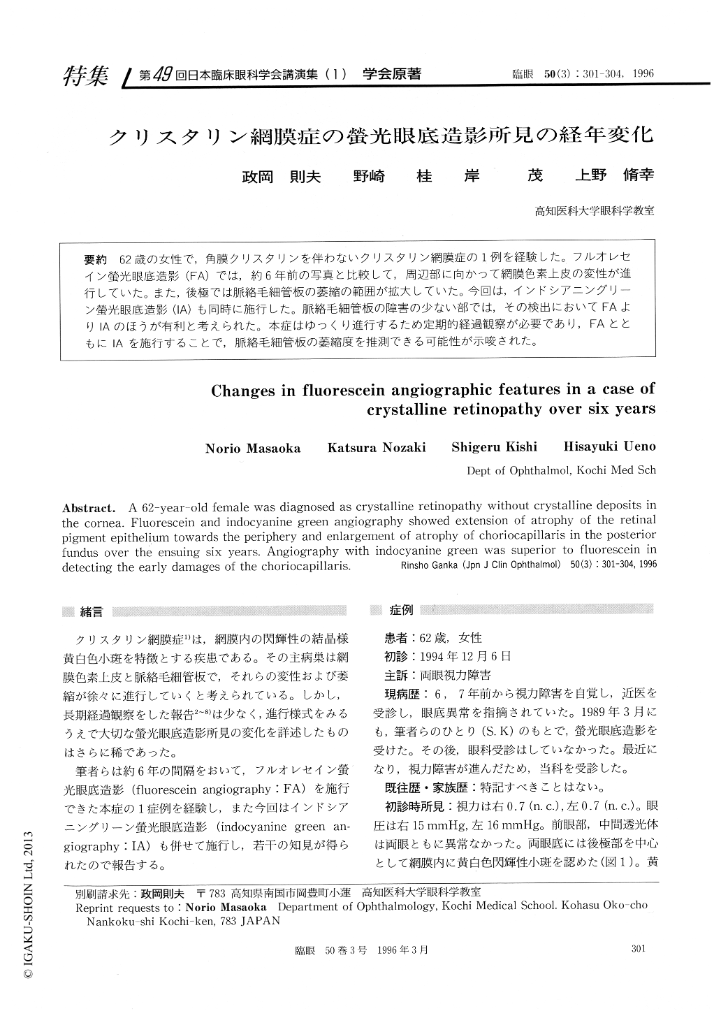
62歳の女性で,角膜クリスタリンを伴わないクリスタリン網膜症の1例を経験した。フルオレセイン螢光眼底造影(FA)では,約6年前の写真と比較して,周辺部に向かって網膜色素上皮の変性が進行していた。また,後極では脈絡毛細管板の萎縮の範囲が拡大していた。今回は,インドシアニングリーン螢光眼底造影(IA)も同時に施行した。脈絡毛細管板の障害の少ない部では,その検出においてFAよりIAのほうが有利と考えられた。本症はゆっくり進行するため定期的経過観察が必要であり,FAとともにIAを施行することで,脈絡毛細管板の萎縮度を推測できる可能性が示唆された。
A 62-year-old female was diagnosed as crystalline retinopathy without crystalline deposits in the cornea. Fluorescein and indocyanine green angiography showed extension of atrophy of the retinal pigment epithelium towards the periphery and enlargement of atrophy of choriocapillaris in the posterioi fundus over the ensuing six years. Angiography with indocyanine green was superior to fluorescein in detecting the early damages of the choriocapillaris.

Copyright © 1996, Igaku-Shoin Ltd. All rights reserved.


