Japanese
English
- 有料閲覧
PPV購入案内
1,320円 (1,200円+税10%) こちらは、847ページから854ページの文献です。購入には医書.jp本会員登録が必要です。
会員登録完了後に改めて上記「購入サイトへ」を選択してください。
または「購入サイトへ」選択後に「購入ログイン」、「新規会員登録」の順に進んでください。
会員登録について詳しくはをご確認ください。
- Abstract 文献概要
- 1ページ目 Look Inside
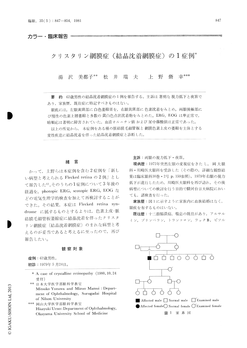
62歳男性の結晶沈着網膜症の1例を報告する。主訴は著明な視力低下と夜盲であり,家族歴,既往症に特記すべきものはない。
眼底には,左眼黄斑部に白色萎縮巣を,右眼黄斑部に色素沈着をみとめ,両眼後極部にび慢性の色素上層萎縮と多数の黄白色点状沈着物をみとめた。ERG,EOGは準正常で,暗順応は著明に障害されていた。血清オルニチン値および尿中蓚酸値は正常であった。
以上の所見から,本症例をある種の脈絡膜毛細管板と網膜色素上皮の萎縮を主体とする変性疾患に結晶沈着を伴った結晶沈着網膜症と診断した。
A 62-year-old male developed severe bilateral decrease of vision and night blindness. Ophthalmo-scopy revealed a white atrophic lesion in the left macula and a pigmented one in the right maculaas well as widespread geographic areas of depig-mentation in the pigment epithelium in both eyes. Numerous, fine, dot-like crystalline deposits were present at the level of the retinal pigment epithelium in both eyes. Fluorescein angiography showed atro-phied retinal pigment epithelium and the chorio-capillaris corresponding to the depigmented areas. The crystalline deposits failed to show any abnormal fluorescein findings.
Copyright © 1981, Igaku-Shoin Ltd. All rights reserved.