Japanese
English
- 有料閲覧
- Abstract 文献概要
- 1ページ目 Look Inside
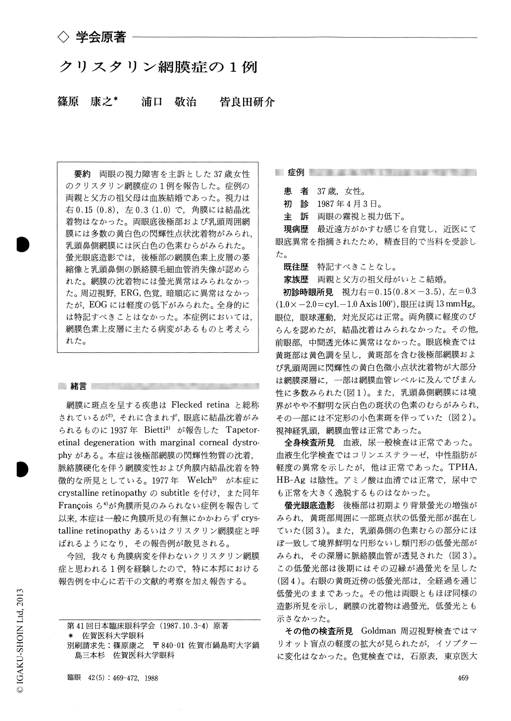
両眼の視力障害を主訴とした37歳女性のクリスタリン網膜症の1例を報告した.症例の両親と父方の祖父母は血族結婚であった.視力は右0.15(0.8),左0.3(1.0)で,角膜には結晶沈着物はなかった.両眼底後極部および乳頭周囲網膜には多数の黄白色の閃輝性点状沈着物がみられ,乳頭鼻側網膜には灰白色の色素むらがみられた.螢光眼底造影では,後極部の網膜色素上皮層の萎縮像と乳頭鼻側の脈絡膜毛細血管消失像が認められた.網膜の沈着物には螢光異常はみられなかった.周辺視野,ERG,色覚,暗順応に異常はなかったが,EOGには軽度の低下がみられた.全身的には特記すべきことはなかった.本症例においては,網膜色素上皮層に主たる病変があるものと考えられた.
A case of crystalline retinopathy was reported. The patient was a 37-year-old female and had visual disturbance in both eyes. Her parents and paternal grandparents were consanguineous. Her corrected visual acuity was RE : 0.8 and LE : 1.0. Slitlamp examination disclosed no abnormality including the cornea. Ophthalmoscopic examina-tion revealed many fine yellowish-white glittering spots scattered mainly in the posterior pole and geographic discoloration of the retina nasal to thedisc in both eyes. Fluorescein angiography showed disorder of the retinal pigment epithelium in the posterior pole and atrophy of the choriocapillaries in the area corresponding with the retinal discolora-tion. Electro-oculography showed subnormality in both eyes, although visual field examination, electroretinography, dark adaptation and color sense were normal. Based on these findings, it was suggested that the main lesion of this case was located in the retinal pigment epithelial layer.
Rinsho Ganka (Jpn J Clin Ophthalmol) 42(5) : 469-472, 1988

Copyright © 1988, Igaku-Shoin Ltd. All rights reserved.


