Japanese
English
- 有料閲覧
PPV購入案内
1,320円 (1,200円+税10%) こちらは、1009ページから1013ページの文献です。購入には医書.jp本会員登録が必要です。
会員登録完了後に改めて上記「購入サイトへ」を選択してください。
または「購入サイトへ」選択後に「購入ログイン」、「新規会員登録」の順に進んでください。
会員登録について詳しくはをご確認ください。
- Abstract 文献概要
- 1ページ目 Look Inside
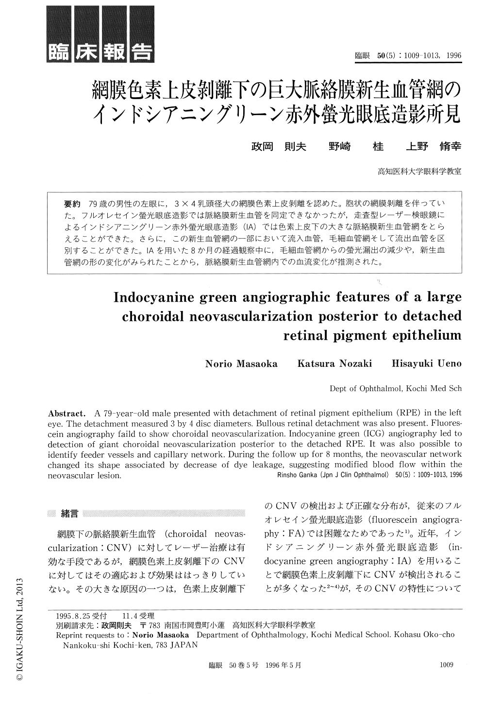
79歳の男性の左眼に,3×4乳頭径大の網膜色素上皮剥離を認めた。胞状の網膜剥離を伴っていた。フルオレセイン螢光眼底造影では脈絡膜新生血管を同定できなかったが,走査型レーザー検眼鏡によるインドシアニングリーン赤外螢光眼底造影(IA)では色素上皮下の大きな脈絡膜新生血管網をとらえることができた。さらに,この新生血管網の一部において流入血管,毛細血管網そして流出血管を区別することができた。IAを用いた8か月の経過観察中に,毛細血管網からの螢光漏出の減少や,新生血管網の形の変化がみられたことから,脈絡膜新生血管網内での血流変化が推測された。
A 79-year-old male presented with detachment of retinal pigment epithelium (RPE) in the left eye. The detachment measured 3 by 4 disc diameters. Bullous retinal detachment was also present. Fluores-cein angiography faild to show choroidal neovascularization. Indocyanine green (ICG) angiography led to detection of giant choroidal neovascularization posterior to the detached RPE. It was also possible to identify feeder vessels and capillary network. During the follow up for 8 months, the neovascular network changed its shape associated by decrease of dye leakage, suggesting modified blood flow within the neovascular lesion.
Copyright © 1996, Igaku-Shoin Ltd. All rights reserved.