Japanese
English
- 有料閲覧
- Abstract 文献概要
- 1ページ目 Look Inside
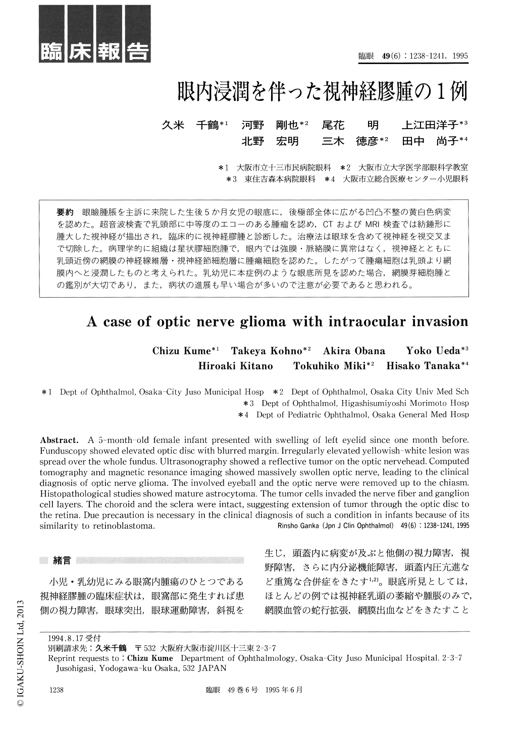
眼瞼腫脹を主訴に来院した生後5か月女児の眼底に,後極部全体に広がる凹凸不整の黄白色病変を認めた。超音波検査で乳頭部に中等度のエコーのある腫瘤を認め,CTおよびMRI検査では紡錘形に腫大した視神経が描出され,臨床的に視神経膠腫と診断した。治療法は眼球を含めて視神経を視交叉まで切除した。病理学的に組織は星状膠細胞腫で,眼内では強膜・脈絡膜に異常はなく,視神経とともに乳頭近傍の網膜の神経線維層・視神経節細胞層に腫瘍細胞を認めた。したがって腫瘍細胞は乳頭より網膜内へと浸潤したものと考えられた。乳幼児に本症例のような眼底所見を認めた場合,網膜芽細胞腫との鑑別が大切であり,また,病状の進展も早い場合が多いので注意が必要であると思われる。
A 5-month-old female infant presented with swelling of left eyelid since one month before. Funduscopy showed elevated optic disc with blurred margin. Irregularly elevated yellowish-white lesion was spread over the whole fundus. Ultrasonography showed a reflective tumor on the optic nervehead. Computed tomography and magnetic resonance imaging showed massively swollen optic nerve, leading to the clinical diagnosis of optic nerve glioma. The involved eyeball and the optic nerve were removed up to the chiasm. Histopathological studies showed mature astrocytoma. The tumor cells invaded the nerve fiber and ganglion cell layers. The choroid and the sclera were intact, suggesting extension of tumor through the optic disc to the retina. Due precaution is necessary in the clinical diagnosis of such a condition in infants because of its similarity to retinoblastoma.

Copyright © 1995, Igaku-Shoin Ltd. All rights reserved.


