Japanese
English
- 有料閲覧
- Abstract 文献概要
- 1ページ目 Look Inside
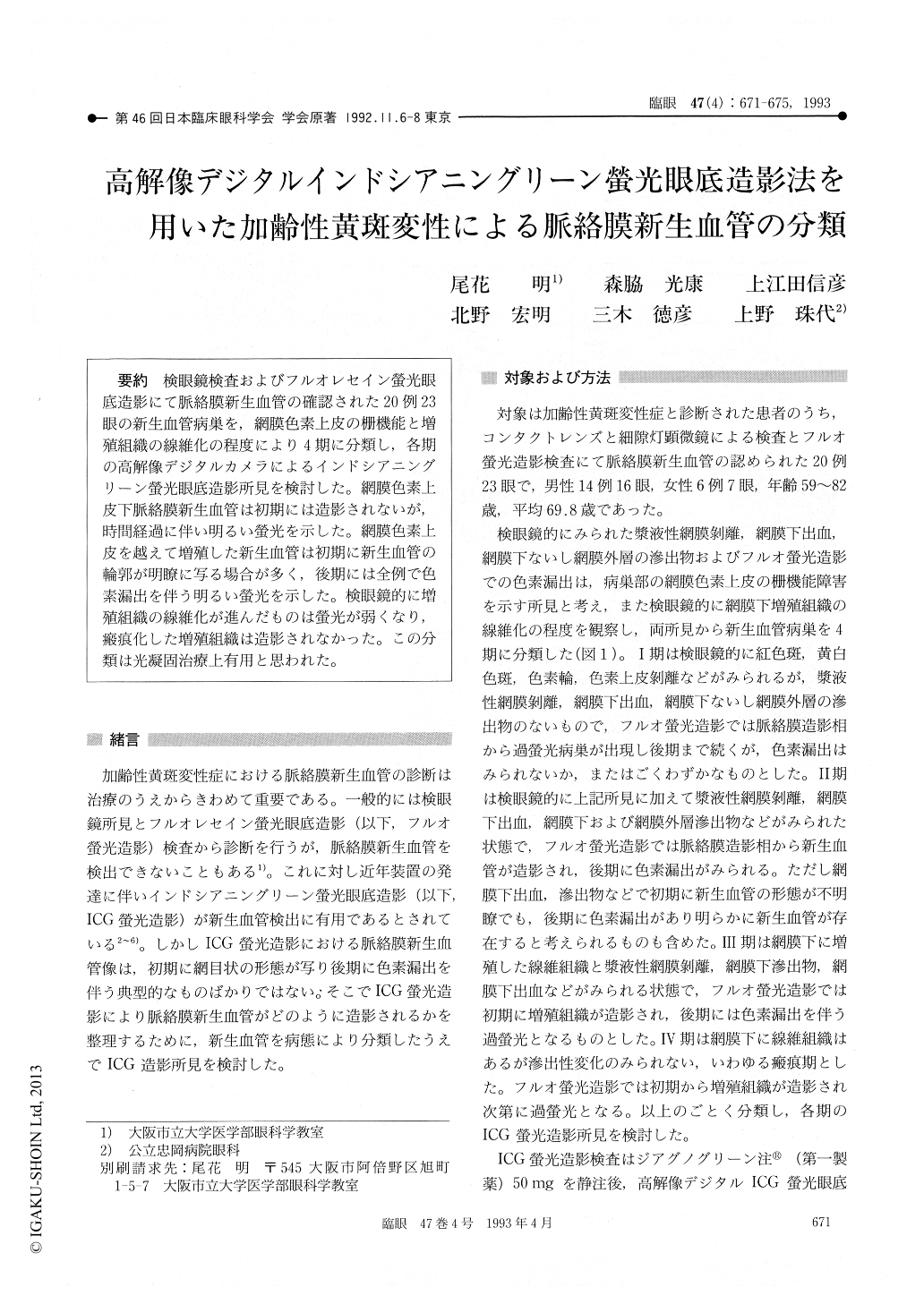
検眼鏡検査およびフルオレセイン螢光眼底造影にて脈絡膜新生血管の確認された20例23眼の新生血管病巣を,網膜色素上皮の柵機能と増殖組織の線維化の程度により4期に分類し,各期の高解像デジタルカメラによるインドシアニングリーン螢光眼底造影所見を検討した。網膜色素上皮下脈絡膜新生血管は初期には造影されないが,時間経過に伴い明るい螢光を示した。網膜色素上皮を越えて増殖した新生血管は初期に新生血管の輪郭が明瞭に写る場合が多く,後期には全例で色素漏出を伴う明るい螢光を示した。検眼鏡的に増殖組織の線維化が進んだものは螢光が弱くなり,瘢痕化した増殖組織は造影されなかった。この分類は光凝固治療上有用と思われた。
We performed high-resolution indocyanine green (ICG) digital angiography in 23 eyes, 20 patients, with age-related macular degeneration. Presence of choroidal neovascularization (CNV) had been con-firmed in all the eyes by ophthalmoscopy and fluor-escein angiography. We could classify these eyes into 4 stages according to the impaired blood -retinal barrier in the retinal pigment epithelium (RPE) and the degree of fibrosis of CNV. CNVs posterior to the RPE failed to be detected in the early-phase ICG angiogram and showed intense fluorescence in the late-phase angiogram. Fluores-cence became less intense along with progression of fibrosis of the CNV. Fibrotic membrane showed no fluorescence. The classification seemed to be of value as a guide for laser treatment.

Copyright © 1993, Igaku-Shoin Ltd. All rights reserved.


