Japanese
English
- 有料閲覧
PPV購入案内
1,320円 (1,200円+税10%) こちらは、611ページから614ページの文献です。購入には医書.jp本会員登録が必要です。
会員登録完了後に改めて上記「購入サイトへ」を選択してください。
または「購入サイトへ」選択後に「購入ログイン」、「新規会員登録」の順に進んでください。
会員登録について詳しくはをご確認ください。
- Abstract 文献概要
- 1ページ目 Look Inside
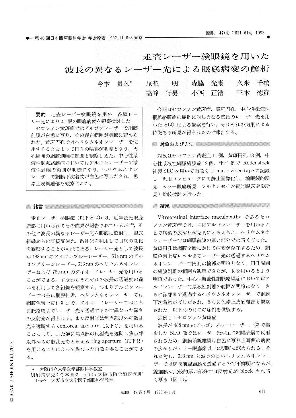
走査レーザー検眼鏡を用い,各種レーザー光により41眼の眼底病変を観察検討した。
セロファン黄斑症ではアルゴンレーザーで網膜前膜が白色に写り,その存在範囲が明瞭に認められた。黄斑円孔ではヘリウムネオンレーザーを使用することによって円孔の輪郭が明瞭となり,円孔周囲の網膜剥離の範囲も観察しえた。中心性漿液性網脈絡膜症においてはアルゴンレーザーで漿液性剥離の範囲が明瞭になり,ヘリウムネオンレーザーで網膜下沈着物が白色に写しだされ,色素上皮剥離部も観察された。
We observed 41 eyes with various fundus diseases by scanning laser ophthalmoscope using argon and helium-neon laser as illumination. Details of pre-retinal membrane in cellophane maculopathy could be observed by argon. Helium-neon was useful in observing the macular hole with surrounding retinal detachment. Argon was useful in observing the margin of detached retina in central serous chorioretinopathy. Subretinal precipitates and detachment of retinal pigment epithelium in this disease could be sharply observed by helium-neon.
Copyright © 1993, Igaku-Shoin Ltd. All rights reserved.