Japanese
English
- 有料閲覧
- Abstract 文献概要
- 1ページ目 Look Inside
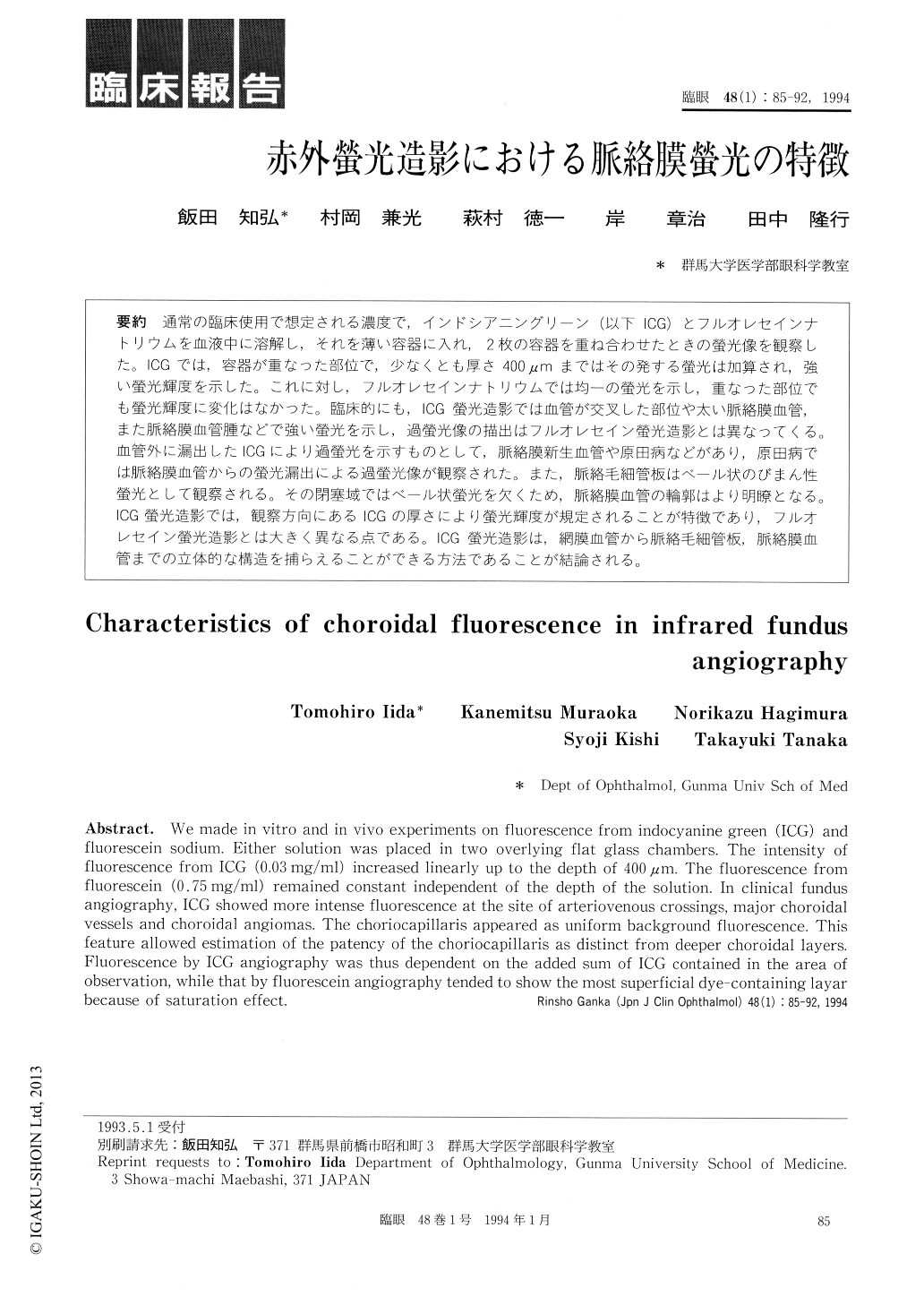
通常の臨床使用で想定される濃度で,インドシアニングリーン(以下ICG)とフルオレセインナトリウムを血液中に溶解し,それを薄い容器に入れ,2枚の容器を重ね合わせたときの螢光像を観察した。ICGでは,容器が重なった部位で,少なくとも厚さ400μmまではその発する螢光は加算され,強い螢光輝度を示した。これに対し,フルオレセインナトリウムでは均一の螢光を示し,重なった部位でも螢光輝度に変化はなかった。臨床的にも,ICG螢光造影では血管が交叉した部位や太い脈絡膜血管,また脈絡膜血管腫などで強い螢光を示し,過螢光像の描出はフルオレセイン螢光造影とは異なってくる。血管外に漏出したICGにより過螢光を示すものとして,脈絡膜新生血管や原田病などがあり,原田病では脈絡膜血管からの螢光漏出による過螢光像が観察された。また,脈絡毛細管板はベール状のびまん性螢光として観察される。その閉塞域ではベール状螢光を欠くため,脈絡膜血管の輪郭はより明瞭となる。ICG螢光造影では,観察方向にあるICGの厚さにより螢光輝度が規定されることが特徴であり,フルオレセイン螢光造影とは大きく異なる点である。ICG螢光造影は,網膜血管から脈絡毛細管板,脈絡膜血管までの立体的な構造を捕らえることができる方法であることが結論される。
We made in vitro and in vivo experiments on fluorescence from indocyanine green (ICG) and fluorescein sodium. Either solution was placed in two overlying flat glass chambers. The intensity of fluorescence from ICG (0.03mg/ml) increased linearly up to the depth of 400μm. The fluorescence from fluorescein (0.75mg/ml) remained constant independent of the depth of the solution. In clinical fundus angiography, ICG showed more intense fluorescence at the site of arteriovenous crossings, major choroidal vessels and choroidal angiomas. The choriocapillaris appeared as uniform background fluorescence. This feature allowed estimation of the patency of the choriocapillaris as distinct from deeper choroidal layers. Fluorescence by ICG angiography was thus dependent on the added sum of ICG contained in the area of observation, while that by fluorescein angiography tended to show the most superficial dye-containing layar because of saturation effect.

Copyright © 1994, Igaku-Shoin Ltd. All rights reserved.


