Japanese
English
- 有料閲覧
- Abstract 文献概要
- 1ページ目 Look Inside
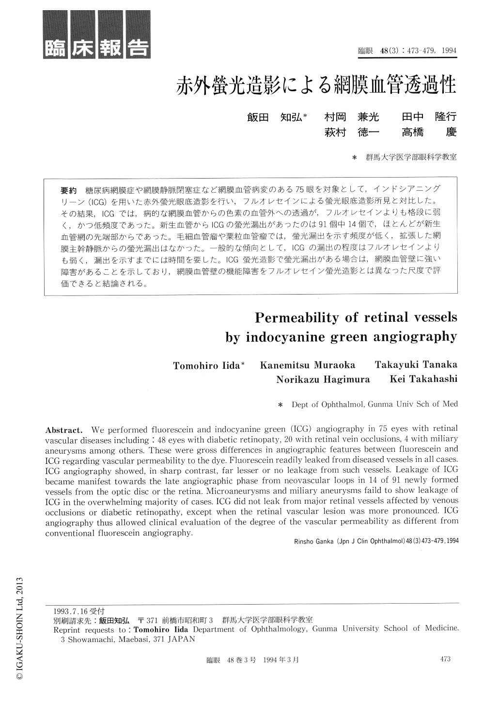
糖尿病網膜症や網膜静脈閉塞症など網膜血管病変のある75眼を対象として,インドシアニングリーン(ICG)を用いた赤外螢光眼底造影を行い,フルオレセインによる螢光眼底造影所見と対比した。その結果,ICGでは,病的な網膜血管からの色素の血管外への透過が,フルオレセインよりも格段に弱く,かつ低頻度であった。新生血管からICGの螢光漏出があったのは91個中14個で,ほとんどが新生血管網の先端部からであった。毛細血管瘤や粟粒血管瘤では,螢光漏出を示す頻度が低く,拡張した網膜主幹静脈からの螢光漏出はなかった。一般的な傾向として,lCGの漏出の程度はフルオレセインよりも弱く,漏出を示すまでには時間を要した。ICG螢光造影で螢光漏出がある場合は,網膜血管壁に強い障害があることを示しており,網膜血管壁の機能障害をフルオレセイン螢光造影とは異なった尺度で評価できると結論される。
We performed fluorescein and indocyanine green (ICG) angiography in 75 eyes with retinal vascular diseases including: 48 eyes with diabetic retinopaty, 20 with retinal vein occlusions, 4 with miliary aneurysms among others. These were gross differences in angiographic features between fluorescein and ICG regarding vascular permeability to the dye. Fluorescein readily leaked from diseased vessels in all cases. ICG angiography showed, in sharp contrast, far lesser or no leakage from such vessels. Leakage of ICG became manifest towards the late angiographic phase from neovascular loops in 14 of 91 newly formed vessels from the optic disc or the retina. Microaneurysms and miliary aneurysms faild to show leakage of ICG in the overwhelming majority of cases. ICG did not leak from major retinal vessels affected by venous occlusions or diabetic retinopathy, except when the retinal vascular lesion was more pronounced. ICG angiography thus allowed clinical evaluation of the degree of the vascular permeability as different from conventional fluorescein angiography.

Copyright © 1994, Igaku-Shoin Ltd. All rights reserved.


