Japanese
English
- 有料閲覧
- Abstract 文献概要
- 1ページ目 Look Inside
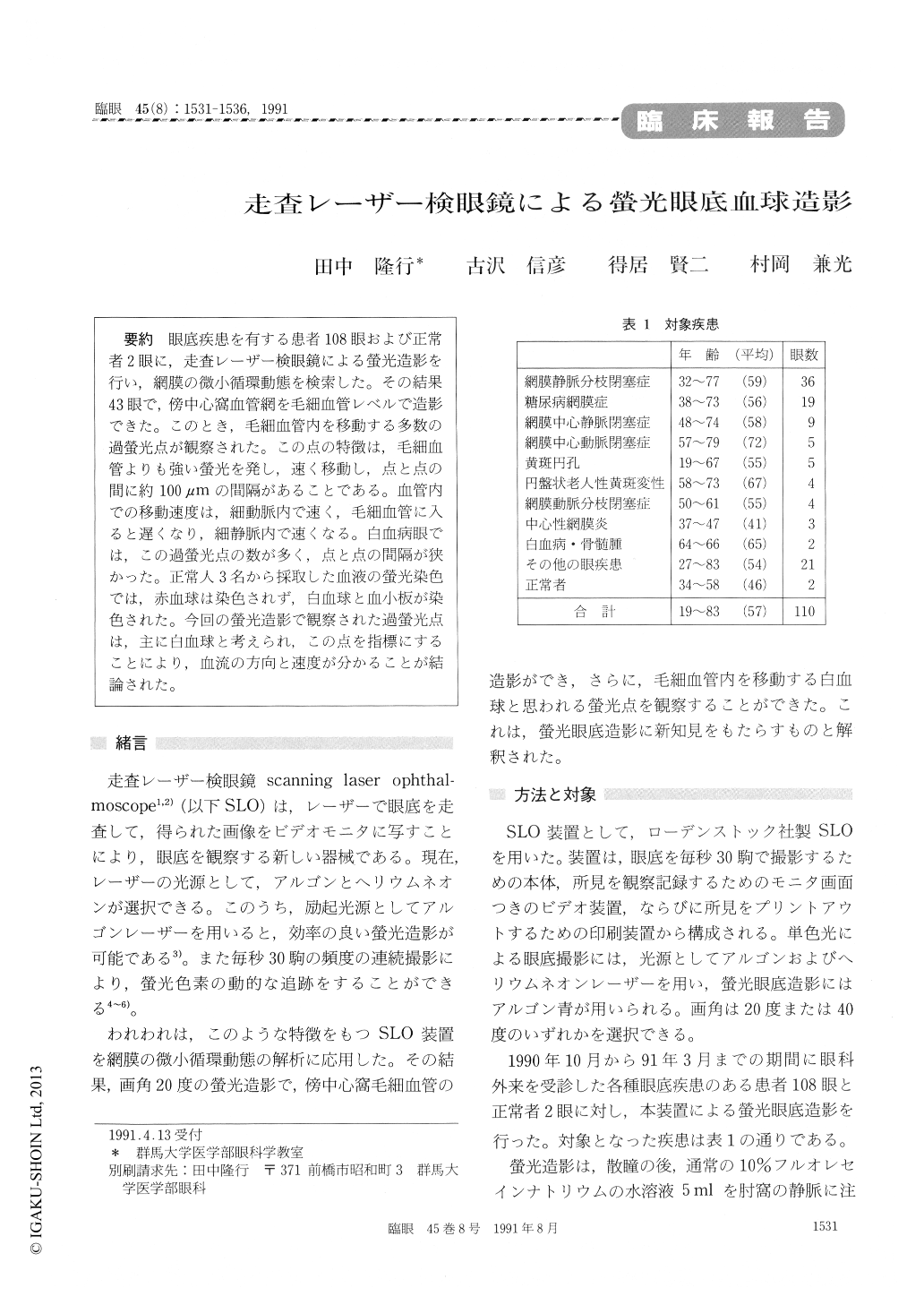
眼底疾患を有する患者108眼および正常者2眼に,走査レーザー検眼鏡による螢光造影を行い,網膜の微小循環動態を検索した。その結果43眼で,傍中心窩血管網を毛細血管レベルで造影できた。このとき,毛細血管内を移動する多数の過螢光点が観察された。この点の特徴は,毛細血管よりも強い螢光を発し,速く移動し,点と点の間に約100μmの間隔があることである。血管内での移動速度は,細動脈内で速く,毛細血管に入ると遅くなり,細静脈内で速くなる。白血病眼では,この過螢光点の数が多く,点と点の間隔が狭かった。正常人3名から採取した血液の螢光染色では,赤血球は染色されず,白血球と血小板が染色された。今回の螢光造影で観察された過螢光点は,主に白血球と考えられ,この点を指標にすることにより,血流の方向と速度が分かることが結論された。
We performed fluorescein fundus angiography in 110 eyes using a scanning laser ophthalmoscope (SLO). We could observe capillaries in the per-ifoveal area in 43 eyes. In all these eyes, as a unique phenomenon, numerous fluorescent dots were seen flowing through the capillaries. The distance from one dot to the next one was widely variable and averaged 100 μm. The velocity of flow was faster in the precapillary arterioles and postcapillaryvenules than in the capillary vessels proper. In a patient with leukemia, the dots were much more numerous than in other, nonleukemic subjects. Staining of whole blood from three healthy persons showed prompt and intense staining of leukocytes and platelets. This finding indicated that the obser-ved fluorescent dots in perifoveal capillaries are mainly leukocytes in the circulating blood. The present findings indicate that fluorescein angiogra-phy with SLO allows identification of the direction and velocity of blood flow in the retinal capillaries.

Copyright © 1991, Igaku-Shoin Ltd. All rights reserved.


