Japanese
English
- 有料閲覧
- Abstract 文献概要
- 1ページ目 Look Inside
緒 言
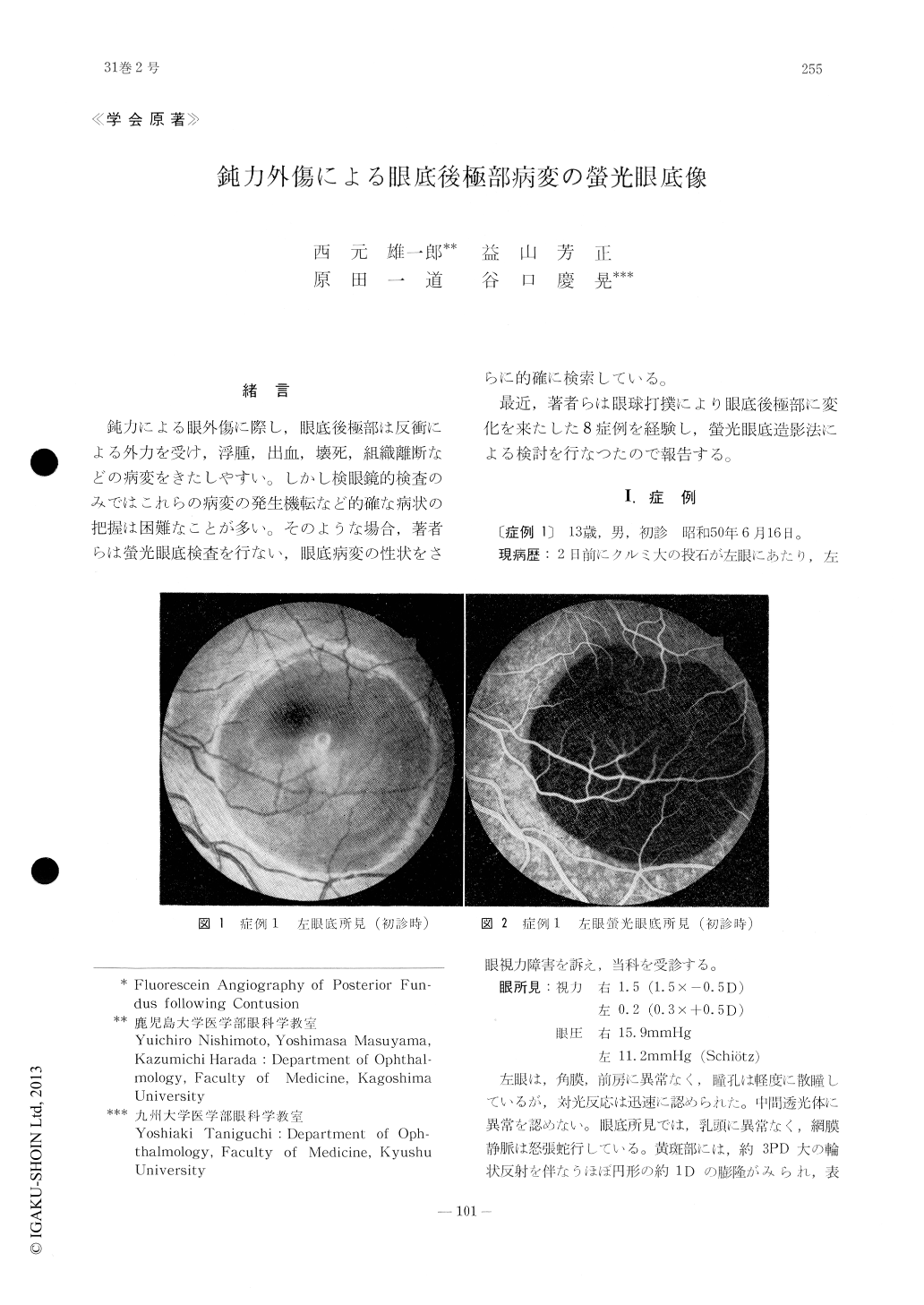
鈍力による眼外傷に際し,眼底後極部は反衝による外力を受け,浮腫,出血,壊死,組織離断などの病変をきたしやすい。しかし検眼鏡的検査のみではこれらの病変の発生機転など的確な病状の把握は困難なことが多い。そのような場合,著者らは螢光眼底検査を行ない,眼底病変の性状をさらに的確に検索している。
最近,著者らは眼球打撲により眼底後極部に変化を来たした8症例を経験し,螢光眼底造影法による検討を行なつたので報告する。
We examined 8 eyes with fundus lesions following blunt contusion with the use of fluo-rescien angiography. Fluorescein angiography facilitated detection of lesions such as subretinal hemorrhage and chorioretinal rupture even when the turbid retina hampered the routine diagnostic examinations. Traumatic macular hole was characterized as small fluorescent patch without a tendency to enlarge in size. Extravasation of fluorescein was consistently absent from the retinal capillaries. We frequen-tly noted a segmental atrophic lesion adjacent to the disc, suggesting the presence of impaired choroidal circulation in this area.

Copyright © 1977, Igaku-Shoin Ltd. All rights reserved.


