Japanese
English
- 有料閲覧
- Abstract 文献概要
- 1ページ目 Look Inside
緒言
Novotny-Alvis (1961年)1)らにより螢光眼底撮影法が開発されて以来,現在では螢光眼底撮影は,種々の眼底病変の検索に欠くことのできない臨床検査法の一つとなつてきた。
現在一般に普及し,使用されている直像式螢光眼底カメラを用いて眼底周辺部の撮影を行なうと,その撮影可能な範囲に限界があり,真の眼底周辺部の螢光眼底撮影は困難である。
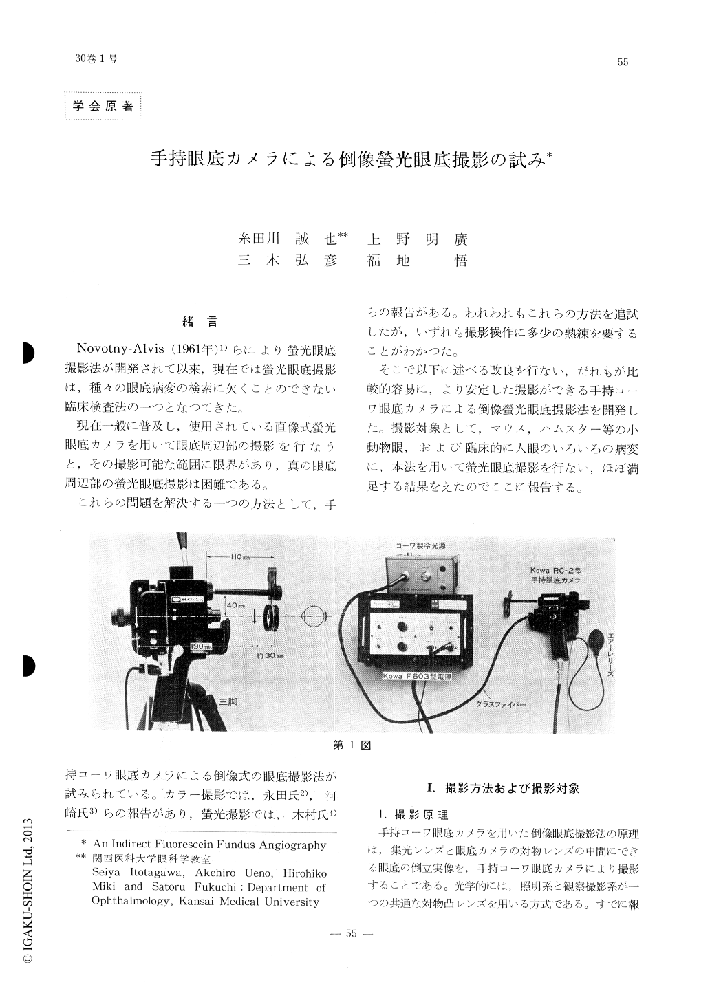
To take fluorescein angiography of the peri-pheral fundus with more ease and certainty, a technique of indirect fundus photography or fluorography using a portable Kowa RC-2 fun-dus camera, as previously described by M. Na-gata and C. Kimura, was further improved by adding new devices. Using this method peri-pheral fundus fluorography was excuted insmall eyes of experimental animals such as mice and hamsters and peripheral lesions of human eyes such as oral disinsertion, immature retinopathy and suspected choraidal melanoma with satisfactory results. Some of them were demonstrated.

Copyright © 1976, Igaku-Shoin Ltd. All rights reserved.


