Japanese
English
- 有料閲覧
- Abstract 文献概要
- 1ページ目 Look Inside
- 参考文献 Reference
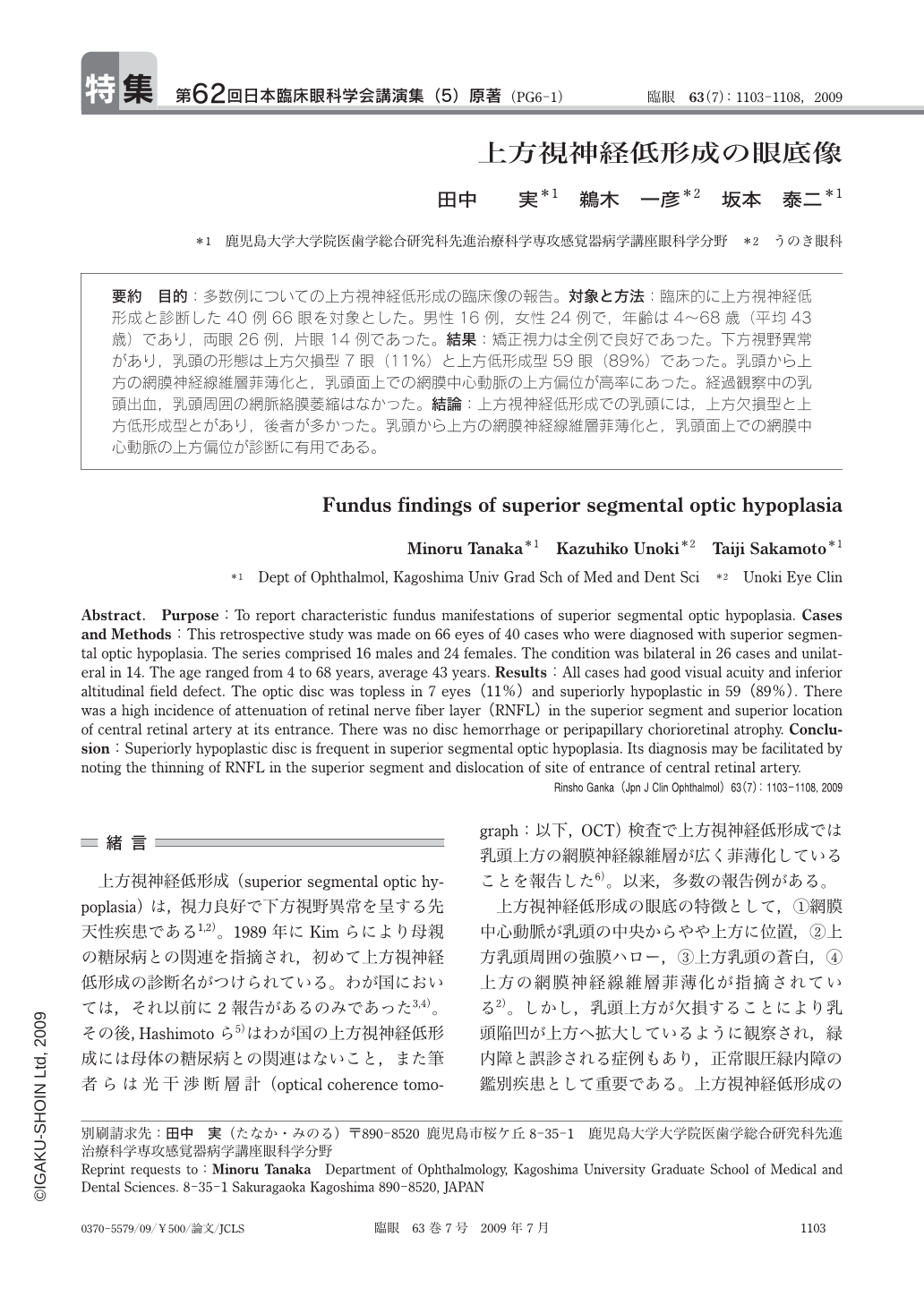
要約 目的:多数例についての上方視神経低形成の臨床像の報告。対象と方法:臨床的に上方視神経低形成と診断した40例66眼を対象とした。男性16例,女性24例で,年齢は4~68歳(平均43歳)であり,両眼26例,片眼14例であった。結果:矯正視力は全例で良好であった。下方視野異常があり,乳頭の形態は上方欠損型7眼(11%)と上方低形成型59眼(89%)であった。乳頭から上方の網膜神経線維層菲薄化と,乳頭面上での網膜中心動脈の上方偏位が高率にあった。経過観察中の乳頭出血,乳頭周囲の網脈絡膜萎縮はなかった。結論:上方視神経低形成での乳頭には,上方欠損型と上方低形成型とがあり,後者が多かった。乳頭から上方の網膜神経線維層菲薄化と,乳頭面上での網膜中心動脈の上方偏位が診断に有用である。
Abstract. Purpose:To report characteristic fundus manifestations of superior segmental optic hypoplasia. Cases and Methods:This retrospective study was made on 66 eyes of 40 cases who were diagnosed with superior segmental optic hypoplasia. The series comprised 16 males and 24 females. The condition was bilateral in 26 cases and unilateral in 14. The age ranged from 4 to 68 years,average 43 years. Results:All cases had good visual acuity and inferior altitudinal field defect. The optic disc was topless in 7 eyes(11%)and superiorly hypoplastic in 59(89%). There was a high incidence of attenuation of retinal nerve fiber layer(RNFL)in the superior segment and superior location of central retinal artery at its entrance. There was no disc hemorrhage or peripapillary chorioretinal atrophy. Conclusion:Superiorly hypoplastic disc is frequent in superior segmental optic hypoplasia. Its diagnosis may be facilitated by noting the thinning of RNFL in the superior segment and dislocation of site of entrance of central retinal artery.
Copyright © 2009, Igaku-Shoin Ltd. All rights reserved.