Japanese
English
- 有料閲覧
- Abstract 文献概要
- 1ページ目 Look Inside
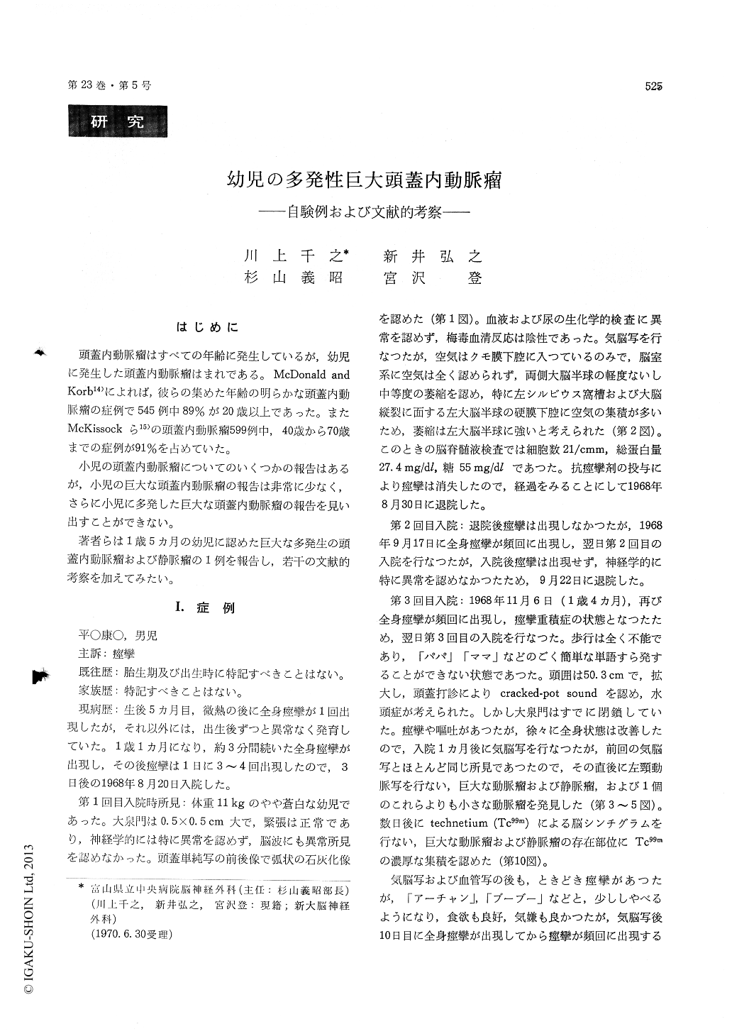
はじめに
頭蓋内動脈瘤はすべての年齢に発生しているが,幼児に発生した頭蓋内動脈瘤はまれである。McDonald andKorb14)によれば,彼らの集めた年齢の明らかな頭蓋内動脈瘤の症例で545例中89%が20歳以上であった。またMcKissockら15)の頭蓋内動脈瘤599例中,40歳から70歳までの症例が91%を占めていた。
小児の頭蓋内動脈瘤についてのいくつかの報告はあるが,小児の巨大な頭蓋内動脈瘤の報告は非常に少なく,さらに小児に多発した巨大な頭蓋内動脈瘤の報告を見い出すことができない。
Although intracranial aneurysms have been re-corded at the extremes of life, they are far less com-mon in infancy and childhood. Several reports of intracranial aneurysms in infants and children have been reported, but no reports of cases of muliple huge intracranial aneurysms in infants and child-ren can be found. Such a case of multiple huge intracranial aneurysms in a young child is reported.
Report of a case : The patient had an attack of general convulsion following slight fever at the age of 5 months, but his development was uneventful until he was 11/12 years old, when several attacks of general convulsion occurred. Neurological ex-amination and electroencephalography revealed no abnormal findings. There was no bulging at the anterior fontanelle, which measured 0. 5 × 0. 5 cm. But the antero-posterior view of the craniogram demonstrated calcification forming a curvilinear line in the left frontal region (Fig. 1), which was later revealed to be calcification in the wall of a huge aneurysm by angiography. Lumbar spinal tap yielded watery clear cerebrospinal fiuld under a normal pressure. Pneumoencephalography revealed marked dilatation of the sylvian fissure on the left side, large amounts of subdural air over the surface of left cerebral hemisphere along the falx cerebri, and no filling of air in the whole ventricular system (Fig. 2). Convulsive seizures were generally con-trolled under the administration of anticonvulsive drugs. At the age of 1 4/12 occurred general con-vulsions followed by status epilepticus. There was enlargement of head circumference measuring 50. 3 cm, and cracked pot sound on percussion of the cranium was obtained. Pneumoencephalography revealed the same findings as those of formerly performed. Left carotid angiography immediately performed after the pneumoencephalography re-vealed a huge and a smaller aneurysms arising fromthe hypertrophied left middle cerebral artery, a huge aneurysm of the left transverse sinus, arterio-venous malformation, and kinking of the left internal carotid artery (Figs. 3-5, 9). Brain scanning with technetium 99m demonstrated the high up-take within the huge aneurysms (Fig. 10). Right carotid angiography revealed marked elevation and bowing of thd anterior cerebral artery, suggesting the presence of hydrocephalus, and hypertrophy of horizontal portions of both anterior cerebral arteries, anterior communicating artery and left middie cerebral artery, which supplied the huge aneurysm, as well as coiling of the right internal carotid artery. Since the age of 1 8/12, he was in the state of akinetic mutism. He expired at the age of 2 5/12. Head had been gradually enlarging and its circumference was 57 cm at the time of death.
Hydrocephalus was thought to occur secondary to adhesive arachnoiditis due to subarachnoid bleed-ing.
Akinetic mutism was considered to be the result of extensive damage to bilateral cerebral white matter caused by increased intracranial pressure secondary to obstructive hydrocephalus.
Seventeen cases of intracranial aneurysms under the age of five years were collected from literat-ures (Table 1).
Out of twelve cases in which the distinction of sex was described, eleven were boys and only one was a girl.
As to the anatomical location, eight arose from the middle cerebral artery, seven from the vertebro-basilar system, one from the anterior cerebral, and one from the internal carotid artery.
Almost all cases had no cranial nerve abnorma lities and exhibited subarachnoid hemorrhage as the initial symptom. Two cases showed signs of spacetaking lesion and one showed cranial nerve abnormalities.
As above described, clinical and pathological features of intracranial aneurysms in infants and young children under the age of five years were found to be greatly different from those in the adults.
In considering the peculiar clinical course of our reported case, it should be pointed out that the cerebral angiography should be performed in cases of infants and young children with uncontrollable epileptic seisures. Curvilinear calcification in plain films of the skull justifies the strong suspicion of the intracranial aneurysm. Establishment of diagno-sis and operation should be done as early as possible.
Copyright © 1971, Igaku-Shoin Ltd. All rights reserved.