Japanese
English
- 有料閲覧
- Abstract 文献概要
- 1ページ目 Look Inside
I.はじめに
先天性頭蓋破裂のうち頭蓋底部脳脱(basal encephal—ocele, anterior naso-frontal encephalocele,intranasalencephalocele)は比較的稀なものである。この頭蓋底部脳脱も脳脱の部位により種々の分類がなされているが,最近著者らは主に神経放射線学的に診断し,手術的に軽快せしめた1例のtranssphenoidal encephaloceleを経験した。本症はまだ本邦で報告がないので,その診断と治療について文献的考察を加え報告する。
Rare malformations are basal encephaloceles (an-terior nasofrontal encephaloceles, intranasal ence-phaloceles) which have been classified as basal and sincipital types. In the basal type, the hernia may be present in the nasopharynx, orbit or mouth, and no external mass is visible. Six forms of the basal type have been classified ; spheno-orbital, sphenomaxillary, sphenoethmoidal, transethmoidal, transsphenoidal, and intrasphenoidal. In the sin-cipital type, an external mass is visible in the nasofrontal region. Sincipital type has been further subdivided into nasofrontal, nasoethmoidal and naso-orbital.
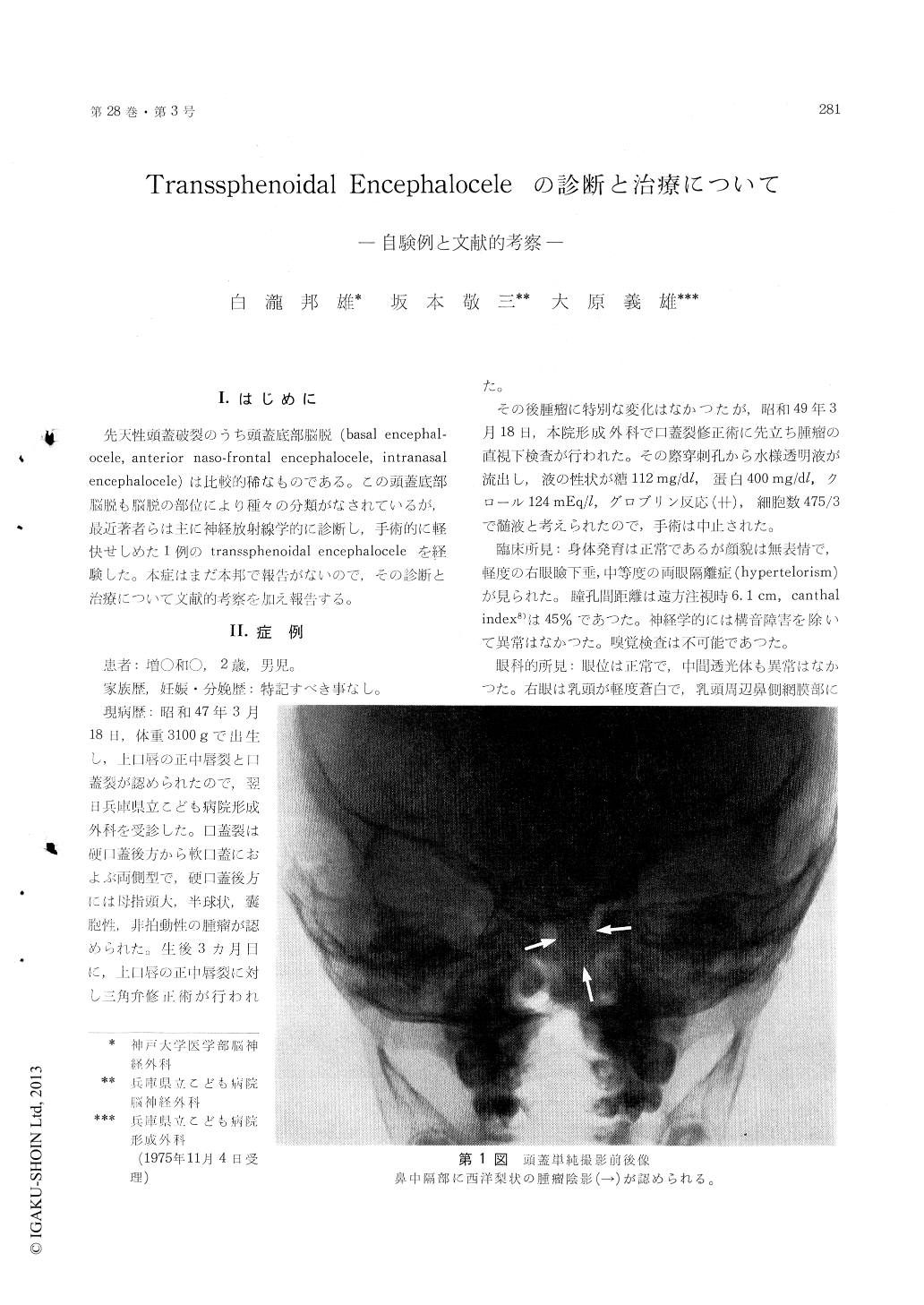
We experienced a case of transspenoidal encephalo-cele in a boy. At birth this boy had a median cleft lip and palate, and a small cystic mass was seen to project through the palatal defect. Mild hypertelorism was also noted. He had the cleft lip repaired surgically at three months of age. When the child was two years old, puncture of the nasopharyngeal mass was done with the result of an escape of a small amount of liquor. Plane roentgenography and tomography of the skull demonstrated a bony defect, 3.3 × 1.5 cm in size, in the floor of the sella, and a soft-tissue mass projecting into the nasopharynx through the defect. A right carotid angiogram revealed downward de-flection of the anterior cerebral artery. A pneumo-encephalogram disclosed downward extension of the prechiasmatic cistern and of the anterior third ven-tricle into the epipharynx. There was also agenesis of the corpus callosum. Exploration of the frontal fossa and the sellar region was carried out through a bifrontal craniotomy exposed by a transcoronal skin incision. Bilateral frontal poles were retracted posteriorly, and downward displacement of the cribriform plate was noted. Further retraction of both rectal gyri showed an anterior part of a bony defect in the sphenoid. All of the displaced struc-tures within the sac could not be returned to the cranial cavity. A pad of Spongel was placed over the evaginated dura mater within the sac.
Up to date twenty-three cases of transsphenoidal encephalocele have been described by twenty re-porters in the literature. In Japan we have found no report on it. From a general viewpoint of eighteen cases including our case, we discussed the clinical features, neurological findings, surgical treatment and pathogenesis of the transsphenoidal encephalocele.
1) Fourteen of the patients were males and three were females, exclusive of one.
2) A congenital nasopharyngeal cystic mass is an important physical finding, suggesting the pres-ence of basal encephalocele. Usually, the mass is encountered at birth through the cleft palate.
3) Both median cleft lip and palate were visualized in seven cases.
4) Moderate hypertelorism is a common feature.
5) In some patients without median cleft palate, the diagnosis was not confirmed until in adult life, although they showed gradually exaggerated chias-matic syndrome. Ocular malformations might be present, such as coloboma, chorioretinal atrophy, unilateral ptosis.
6) The importance of neuroradiologic techniques in diagnosis of this rare lesion has been already stressed by Pollock et al.
7) Including our case, twelve of the eighteen patients were operated on. External ligation or amputation of the pedicle resulted in fetal posto-perative course in three cases. Intracranial route was employed in seven cases, with two postoperative deaths. On the other hand, two transpalatal cor-rections were reported.
Copyright © 1976, Igaku-Shoin Ltd. All rights reserved.