Japanese
English
- 有料閲覧
PPV購入案内
1,650円 (1,500円+税10%) こちらは、583ページから592ページの文献です。購入には医書.jp本会員登録が必要です。
会員登録完了後に改めて上記「購入サイトへ」を選択してください。
または「購入サイトへ」選択後に「購入ログイン」、「新規会員登録」の順に進んでください。
会員登録について詳しくはをご確認ください。
- Abstract 文献概要
- 1ページ目 Look Inside
- 参考文献 Reference
- サイト内被引用 Cited by
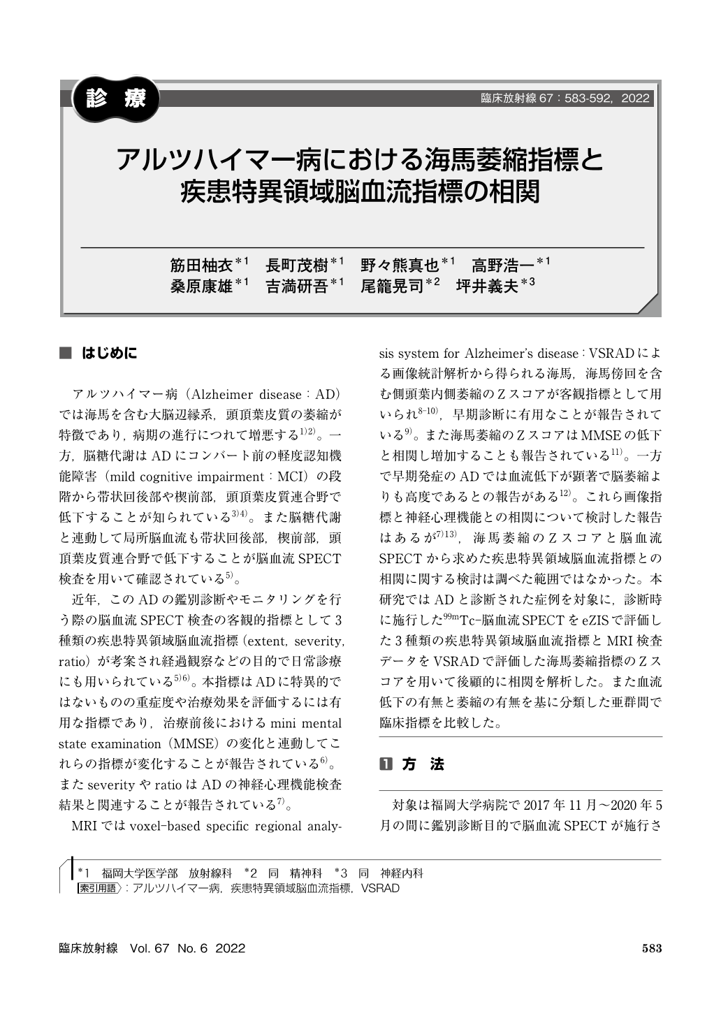
アルツハイマー病(Alzheimer disease:AD)では海馬を含む大脳辺縁系,頭頂葉皮質の萎縮が特徴であり,病期の進行につれて増悪する1)2)。一方,脳糖代謝はADにコンバート前の軽度認知機能障害(mild cognitive impairment:MCI)の段階から帯状回後部や楔前部,頭頂葉皮質連合野で低下することが知られている3)4)。また脳糖代謝と連動して局所脳血流も帯状回後部,楔前部,頭頂葉皮質連合野で低下することが脳血流SPECT検査を用いて確認されている5)。
We examined retrospectively rCBF SPECT and MRI performed at the time of diagnosis in 82 patients diagnosed with Alzheimer’s disease(AD). The hippocampal atrophy was defined as the case with Z score of 2 or more by MRI and VSRAD analysis, and the decreased blood flow cases was defined with one or more disease-specific region indexes by SPECT were positive. They were classified into subgroups with atrophy-only group(18 cases), low perfusion only group(17 cases), both atrophy and low perfusion group(33 cases), and the group without any atrophy and without abnormal perfusion(14 cases). In analyzing among all the cases, no significant correlation was found between the Z-score of hippocampal atrophy and the disease-specific region parameters. In the comparison of HDS-R among subgroups, the both atrophy and hypoperfusion group showed significantly lower values than that of the atrophy-only group or most of hypoperfusion-only group. In the group with both decreased blood flow and atrophy, the decrease in HDS-R was remarkable, but in the group with only either abnormality, the decrease in HDS-R was not so serious. Therefore, in diagnosing AD, imaging study by both SPECT and MRI examinations should be evaluated, even if the decrease of HSR-R is not so severe.
Copyright © 2022, KANEHARA SHUPPAN Co.LTD. All rights reserved.