Japanese
English
- 有料閲覧
- Abstract 文献概要
- 1ページ目 Look Inside
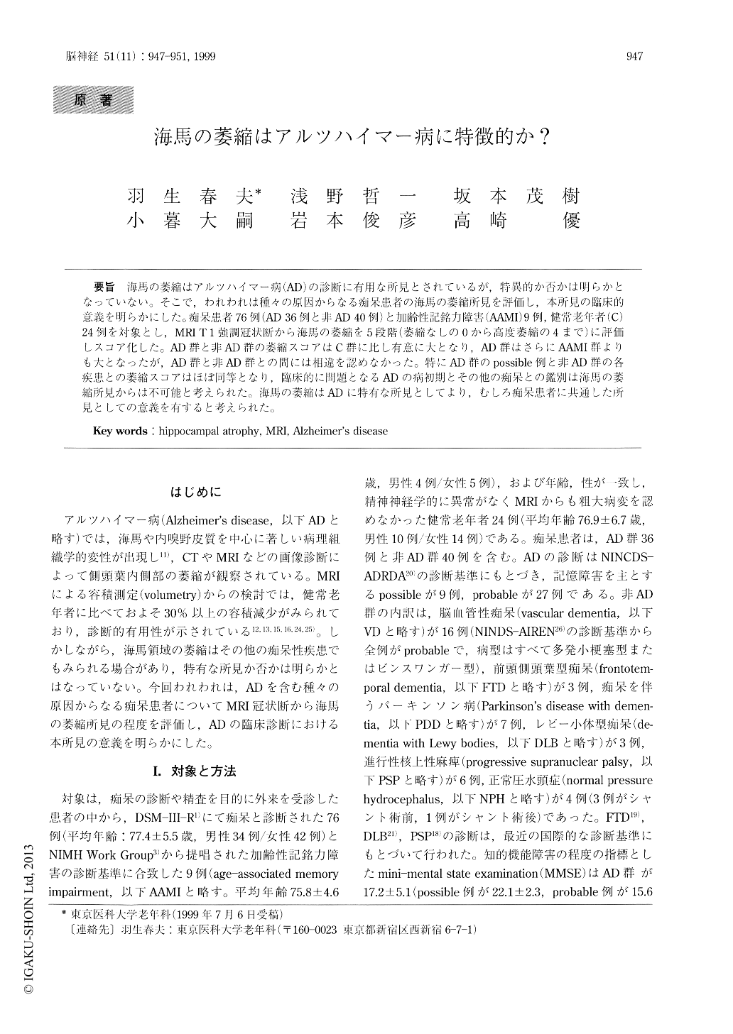
海馬の萎縮はアルツハイマー病(AD)の診断に有用な所見とされているが,特異的か否かは明らかとなっていない。そこで,われわれは種々の原因からなる痴呆患者の海馬の萎縮所見を評価し,本所見の臨床的意義を明らかにした。痴呆患者76例(AD 36例と非AD 40例)と加齢性記銘力障害(AAMI)9例,健常老年者(C)24例を対象とし,MRI T 1強調冠状断から海馬の萎縮を5段階(萎縮なしの0から高度萎縮の4まで)に評価しスコア化した。AD群と非AD群の萎縮スコアはC群に比し有意に大となり,AD群はさらにAAMI群よりも大となったが,AD群と非AD群との間には相違を認めなかった。特にAD群のpossible例と非AD群の各疾患との萎縮スコアはほぼ同等となり,臨床的に問題となるADの病初期とその他の痴呆との鑑別は海馬の萎縮所見からは不可能と考えられた。海馬の萎縮はADに特有な所見としてより,むしろ痴呆患者に共通した所見としての意義を有すると考えられた。
Although detection of hippocampal atrophy has been proposed for the diagnosis of Alzheimer's dis-ease (AD), atrophic changes in MRI can be found in other dementia diseases. This study was undertaken to determine whether hippocampal atrophy was a spe-cific change for AD. Coronal T 1-weighted images were performed in 36 patients with AD, 40 patients with non-AD including vascular dementia, frontempo-ral dementia, Parkinson's disease with dementia, de-mentia with Lewy bodies, progressive supranuclear palsy, and normal pressure hydrocephalus, 9 patients with age-associated memory impairment (AAMI), and 24 control subjects.

Copyright © 1999, Igaku-Shoin Ltd. All rights reserved.


