Japanese
English
- 有料閲覧
PPV購入案内
1,320円 (1,200円+税10%) こちらは、379ページから382ページの文献です。購入には医書.jp本会員登録が必要です。
会員登録完了後に改めて上記「購入サイトへ」を選択してください。
または「購入サイトへ」選択後に「購入ログイン」、「新規会員登録」の順に進んでください。
会員登録について詳しくはをご確認ください。
- Abstract 文献概要
- 1ページ目 Look Inside
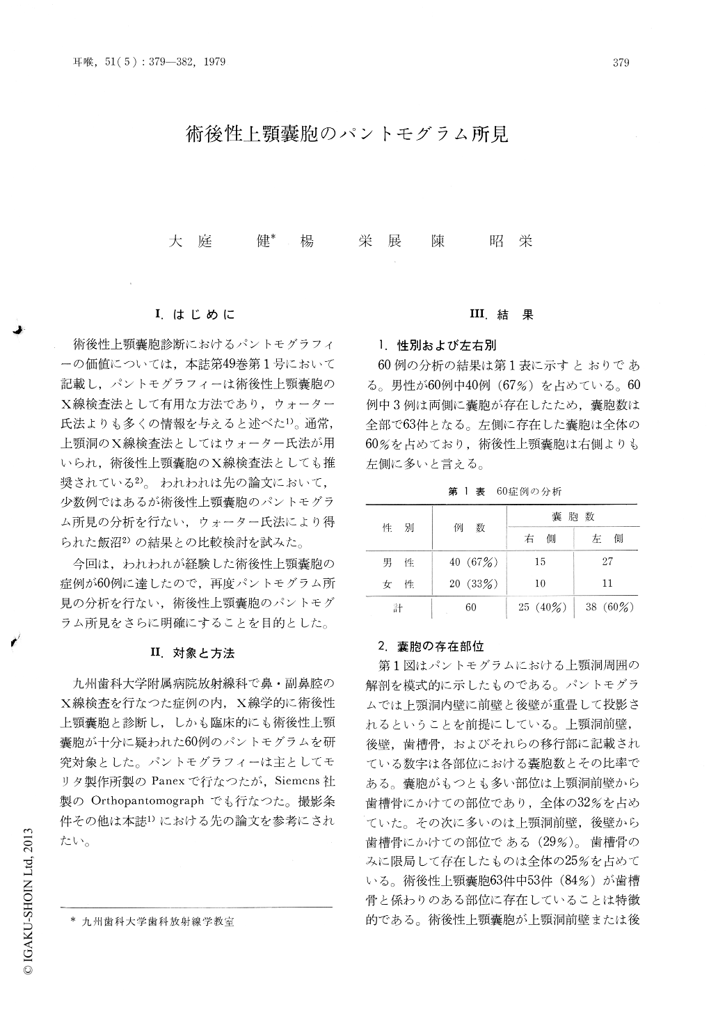
I.はじめに
術後性上顎嚢胞診断におけるパントモグラフィーの価値については,本誌第49巻第1号において記載し,パントモグラフィーは術後性上顎嚢胞のX線検査法として有用な方法であり,ウォーター氏法よりも多くの情報を与えると述べた1)。通常,上顎洞のX線検査法としてはウォーター氏法が用いられ,術後性上顎嚢胞のX線検査法としても推奨されている2)。われわれは先の論文において,少数例ではあるが術後性上顎嚢胞のパントモグラム所見の分析を行ない,ウォーター氏法により得られた飯沼2)の結果との比較検討を試みた。
今回は,われわれが経験した術後性上顎嚢胞の症例が60例に達したので,再度パントモグラム所見の分析を行ない,術後性上顎嚢胞のパントモグラム所見をさらに明確にすることを目的とした。
Sixty cases of postoperative maxillary cyst were studied radiologically by pantomogram. The post-operative maxillary cysts occured more frequently in the male (63%) than in the females, and the left side was more affected (60%) than in the right.
The most characteristic pantomographic findings are a mono-lobular radiolucency accompanied with a well-defined and smooth margin in the anterior-posterior or alveolar region of the maxillay sinus. Marginal sclerosis of the cystic radio-lucency and root resorption are not common in the pantomographic findings of postoperative maxillary cyst.
Copyright © 1979, Igaku-Shoin Ltd. All rights reserved.