Japanese
English
- 有料閲覧
PPV購入案内
1,320円 (1,200円+税10%) こちらは、455ページから462ページの文献です。購入には医書.jp本会員登録が必要です。
会員登録完了後に改めて上記「購入サイトへ」を選択してください。
または「購入サイトへ」選択後に「購入ログイン」、「新規会員登録」の順に進んでください。
会員登録について詳しくはをご確認ください。
- Abstract 文献概要
- 1ページ目 Look Inside
I.はじめに
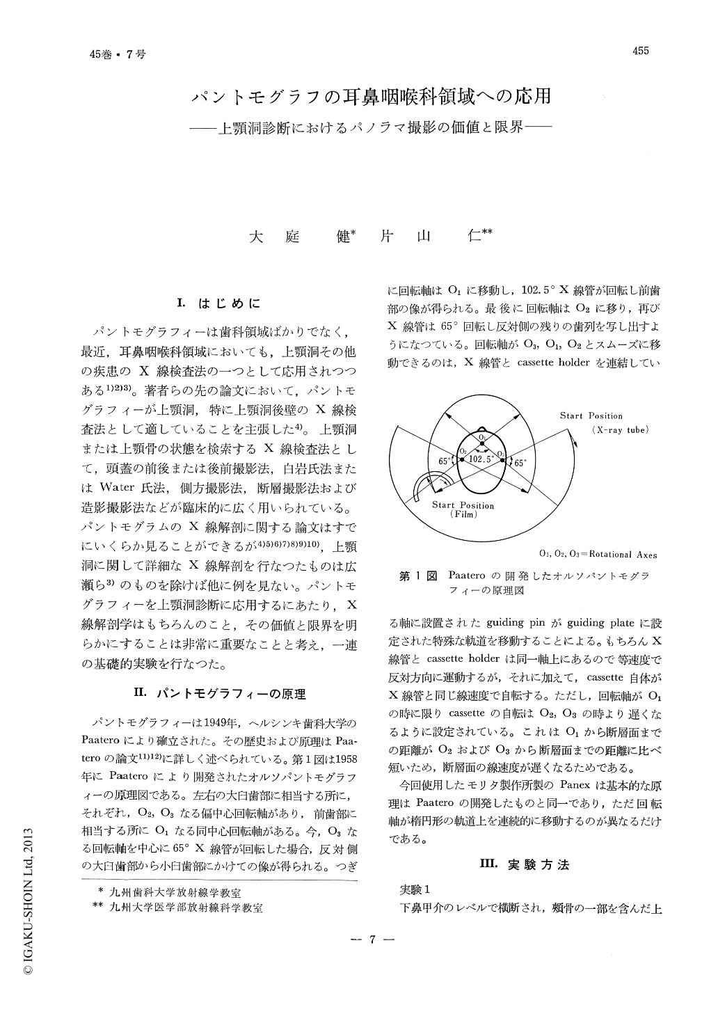
パントモグラフィーは歯科領域ばかりでなく,最近,耳鼻咽喉科領域においても,上顎洞その他の疾患のX線検査法の一つとして応用されつつある1)2)3)。著者らの先の論文において,パントモグラフィーが上顎洞,特に上顎洞後壁のX線検査法として適していることを主張した4)。上顎洞または上顎骨の状態を検索するX線検査法として,頭蓋の前後または後前撮影法,白岩氏法またはWater氏法,側方撮影法,断層撮影法および造影撮影法などが臨床的に広く用いられている。パントモグラムのX線解剖に関する論文はすでにいくらか見ることができるが4)5)6)7)8)9)10),上顎洞に関して詳細なX線解剖を行なつたものは広瀬ら3)のものを除けば他に例を見ない。パントモグラフィーを上顎洞診断に応用するにあたり,X線解剖学はもちろんのこと,その価値と限界を明らかにすることは非常に重要なことと考え,一連の基礎的実験を行なつた。
In order to apply pantomography to radiological diagnosis of the maxillary lesions it is very important not only to know roentgenographic anatomy but also to know value limitation of pantomography.
Lead foils and lines are attached to anatomic landmarks of the dry maxillozygoma specimen and x-ray pictures are taken with Panex, Morita Co., Ltd.
On the pantomogram the medial border of the maxillary sinus is composed of the angle of anterior and medial walls. The lateral border is composed of maximum convexity of the posterior wall.
The anterior, posterior and medial walls are recognized as anatomical landmarks only when these walls are covered by radiopaque material. Most of the anterior and posterior walls in these pictures are superimposed to the medial wall. The anterior wall will appears to occupy medial two thirds of the maxillary sinus and the posterior wall occupies lateral third.
The anterior, posterior and medial walls of the sinus cavity do not appear as definite landmarks on the pantomogram. Moreover, the individual wall is superimposed to each other. Therefore, careful interpretation of the pantomogram is highly essential, for assessment of the maxillary bone destruction.
Copyright © 1973, Igaku-Shoin Ltd. All rights reserved.