Japanese
English
- 有料閲覧
- Abstract 文献概要
- 1ページ目 Look Inside
I.はじめに
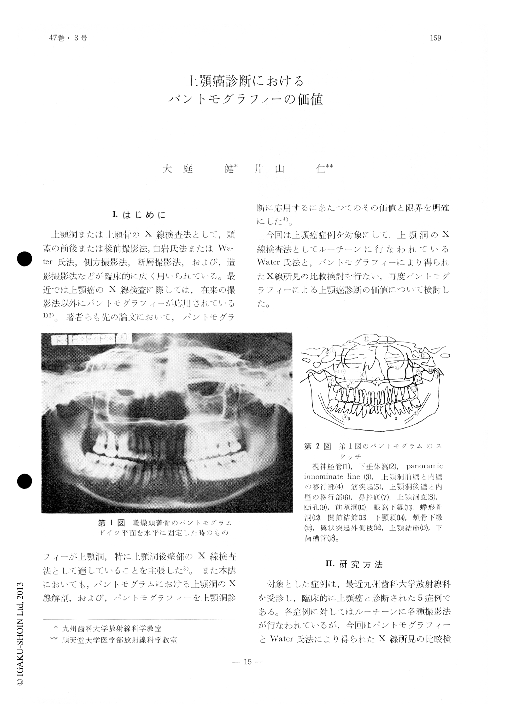
上顎洞または上顎骨のX線検査法として,頭蓋の前後または後前撮影法,白岩氏法またはWater氏法,側方撮影法,断層撮影法,および,造影撮影法などが臨床的に広く用いられている。最近では上顎癌のX線検査に際しては,在来の撮影法以外にパントモグラフィーが応用されている1)2)。著者らも先の論文において,パントモグラフィーが上顎洞,特に上顎洞後壁部のX線検査法として適していることを主張した3)。また本誌においても,パントモグラムにおける上顎洞のX線解剖,および,パントモグラフィーを上顎洞診断に応用するにあたつてのその価値と限界を明確にした4)。
今回は上顎癌症例を対象にして,上顎洞のX線検査法としてルーチーンに行なわれているWater氏法と,パントモグラフィーにより得られたX線所見の比較検討を行ない,再度パントモグラフィーによる上顎癌診断の価値について検討した。
For the diagnosis of cancers in the maxillary sinus a study is made in the use of pantomography with that of routine x-ray examination with Water's method.
Pantomography provides excellent views, not only, of the posterior wall of the maxillary sinus but also, of the conditions in the region of the alveolar bone and the sinus floor whether or not any bony destructions may occur.
The authors emphasize the use of pantomography in combination with other routine x-ray examination would provide important means by which diagnosis of the maxillary sinus pathosis may be made.

Copyright © 1975, Igaku-Shoin Ltd. All rights reserved.


