Japanese
English
- 有料閲覧
PPV購入案内
1,320円 (1,200円+税10%) こちらは、195ページから199ページの文献です。購入には医書.jp本会員登録が必要です。
会員登録完了後に改めて上記「購入サイトへ」を選択してください。
または「購入サイトへ」選択後に「購入ログイン」、「新規会員登録」の順に進んでください。
会員登録について詳しくはをご確認ください。
- Abstract 文献概要
- 1ページ目 Look Inside
I.はじめに
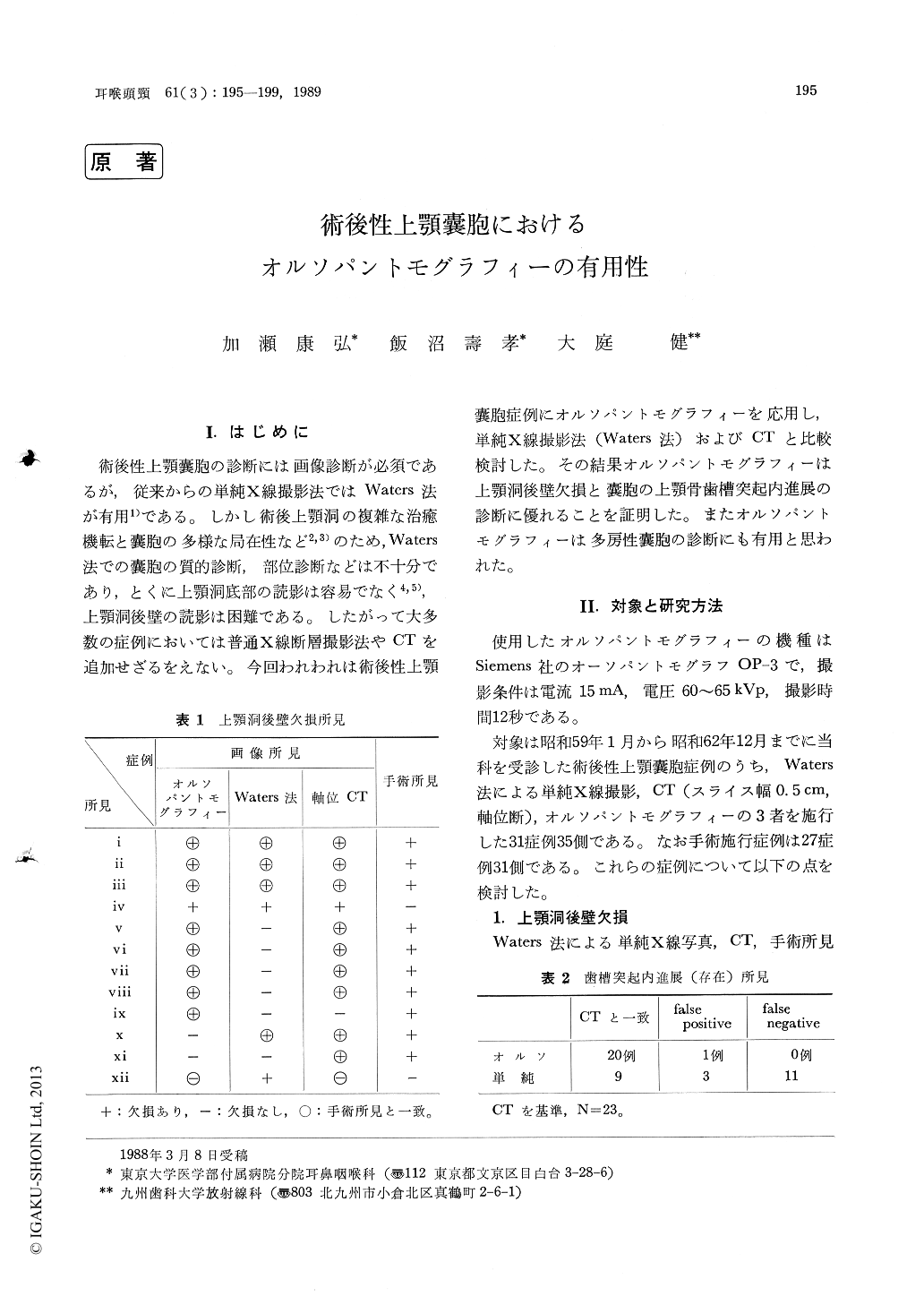
術後性上顎嚢胞の診断には画像診断が必須であるが,従来からの単純X線撮影法ではWaters法が有用1)である。しかし術後上顎洞の複雑な治癒機転と嚢胞の多様な局在性など2,3)のため,Waters法での嚢胞の質的診断,部位診断などは不十分であり,とくに上顎洞底部の読影は容易でなく4,5),上顎洞後壁の読影は困難である。したがって大多数の症例においては普通X線断層撮影法やCTを追加せざるをえない。今回われわれは術後性上顎嚢胞症例にオルソパントモグラフィーを応用し,単純X線撮影法(Waters法)およびCTと比較検討した。その結果オルソパントモグラフィーは上顎洞後壁欠損と嚢胞の上顎骨歯槽突起内進展の診断に優れることを証明した。またオルソパントモグラフィーは多房性嚢胞の診断にも有用と思われた。
Thirty one cases (35 sides) of postoperative cysts of the maxilla were pre-operatively evaluated with conventional x-ray projection (Waters' view), orthopantomography and CT scan (in axial plane). Such findings on images as destruction of the posterior sinus wall and extension of the cyst into the alveolar process were observed on Waters' view, orthopantomography, CT, and these were compared with the postoperative findings. Orthopantomography and CT were equally valuable in evaluating both findings as menticned above, whereas Waters' view is less valuable. In multiple cysts, and cysts located vertically orthopantomography was more valuable than CT in demonstrating anatomical relation-ship of the cysts. Similarly,when the cysts located sagittally orthopantomography was more useful than Waters' view.
Copyright © 1989, Igaku-Shoin Ltd. All rights reserved.