Japanese
English
- 有料閲覧
PPV購入案内
1,320円 (1,200円+税10%) こちらは、41ページから46ページの文献です。購入には医書.jp本会員登録が必要です。
会員登録完了後に改めて上記「購入サイトへ」を選択してください。
または「購入サイトへ」選択後に「購入ログイン」、「新規会員登録」の順に進んでください。
会員登録について詳しくはをご確認ください。
- Abstract 文献概要
- 1ページ目 Look Inside
Ⅰ.はじめに
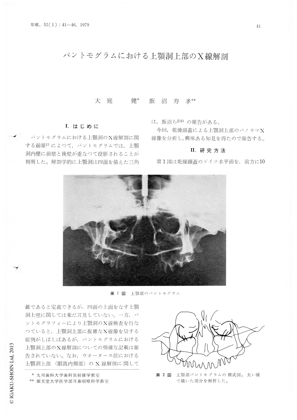
パントモグラムにおける上顎洞のX線解剖に関する前報1)によつて,パントモグラムでは,上顎洞内壁に前壁と後壁が重なつて投影されることが判明した。解剖学的に手上顎洞は四面を備えた三角錐であると定義できるが,四面の上面をなす上顎洞上壁に関しては未だ言及していない。一方,パントモグラフィーにより上顎洞のX線検査を行なつていると,上顎洞上部に複雑なX線像を呈する症例がしばしばあるが,パントモグラムにおける上顎洞上部のX線解剖についての明確な記載は報告されていない。なお,ウオータース位における上顎洞上部(眼窩内側部)のX線解剖に関しては,飯沼ら2)3)の報告がある。
今回,乾燥頭蓋による上顎洞上部のパノラマX線像を分析し,興味ある知見を得たので報告する。
This study was performed to clearify the anatomy of the linear radiopacities which are some-times seen in the superior region of the maxillary sinus on pantomograms. Lead lines were attached on along the sutura frontolacrimalis, sutura lacrimomaxillaris, sutura ethmolacrimalis, crista lacrimalis anterior, crista lacrimalis posterior, medio-superior margin of process uncinatus (inferior margin of hiatus semilunalis), and the superior margin of concha nasalis inferior. Pantomography was performed with the Panex manufactured by the Morita Corporation.
Two major linear radiopacities in the superior region of the maxillary sinus are composed ofthe medio-superior margin of the process uncinatus (inferior margin of hiatus semilunalis) and the superior margin of the concha nasalis inferior.Both nasolacrimal and infraorbital canals are also clearly seen on the superior region of the maxillary sinus on pantomogram.
Copyright © 1980, Igaku-Shoin Ltd. All rights reserved.