Japanese
English
- 有料閲覧
PPV購入案内
1,320円 (1,200円+税10%) こちらは、345ページから350ページの文献です。購入には医書.jp本会員登録が必要です。
会員登録完了後に改めて上記「購入サイトへ」を選択してください。
または「購入サイトへ」選択後に「購入ログイン」、「新規会員登録」の順に進んでください。
会員登録について詳しくはをご確認ください。
- Abstract 文献概要
- 1ページ目 Look Inside
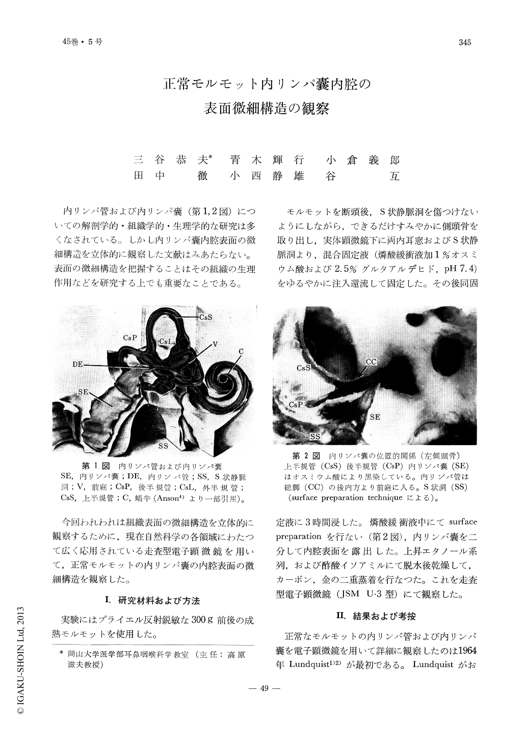
内リンパ管および内リンパ嚢(第1,2図)についての解剖学的・組織学的・生理学的な研究は多くなされている。しかし内リンパ嚢内腔表面の微細構造を立体的に観察した文献はみあたらない。表面の微細構造を把握することはその組織の生理作用などを研究する上でも重要なことである。
今回われわれは組織表面の微細構造を立体的に観察するために,現在自然科学の各領域にわたつて広く応用されている走査型電子顕微鏡を用いて,正常モルモットの内リンパ嚢の内腔表面の微細構造を観察した。
Three dimentional observations of the luminal surface structures of the endolymphatic sac in normal guinea pig were made under the scanning electron microscope.
Based on surface morphology, the endolymphatic sac may be divided into three portions. The proximal portion consists of duct-type cells with short microvilli. The intermediate portion is composed of two kind of cells; cells with closely massed, long microvilli and the other with short microvilli. These cells forming papillae are irregularly arranged. The distal portion is provided with two kinds of cells as seen in the intermediate portion, however, the number and length of the microvilli and the cells are rather sparsely distributed.
Judging from morphological observations of the surface structures, it might be safe to say, functionally, the intermediate portion would be the most active.
Copyright © 1973, Igaku-Shoin Ltd. All rights reserved.