Japanese
English
- 有料閲覧
PPV購入案内
1,320円 (1,200円+税10%) こちらは、697ページから701ページの文献です。購入には医書.jp本会員登録が必要です。
会員登録完了後に改めて上記「購入サイトへ」を選択してください。
または「購入サイトへ」選択後に「購入ログイン」、「新規会員登録」の順に進んでください。
会員登録について詳しくはをご確認ください。
- Abstract 文献概要
- 1ページ目 Look Inside
I.はじめに
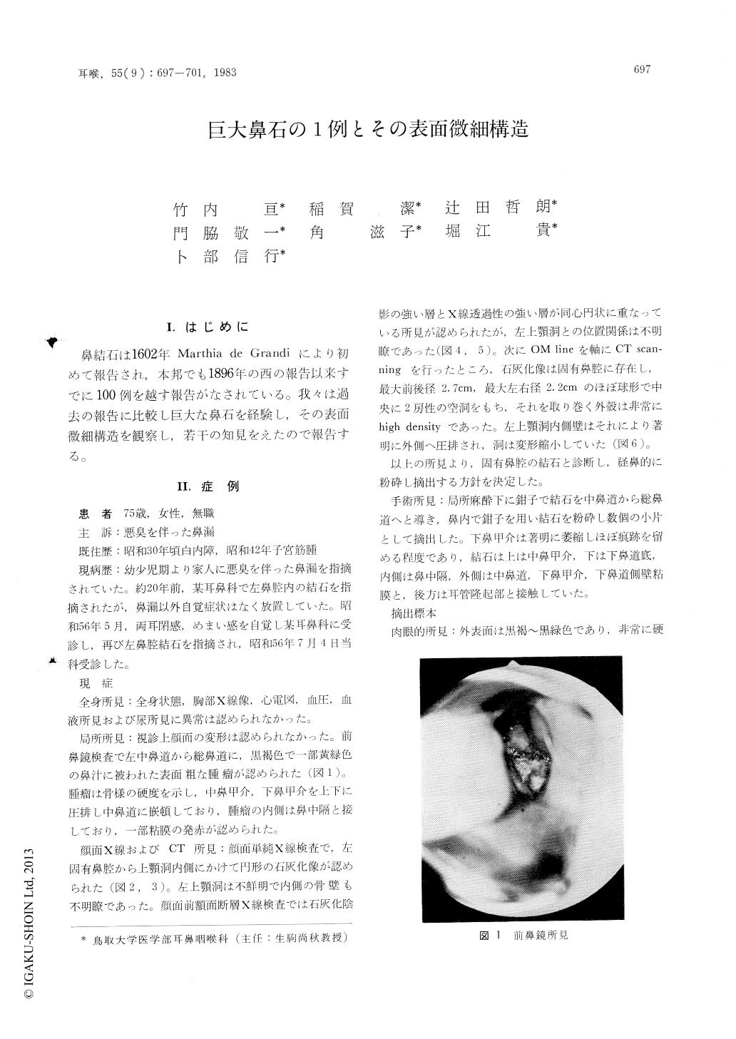
鼻結石は1602年Marthia de Grandiにより初めて報告され,本邦でも1896年の西の報告以来すでに100例を越す報告がなされている。我々は過去の報告に比較し巨大な鼻石を経験し,その表面微細構造を観察し,若干の知見をえたので報告する。
A case of rhinolith is reported. The patient is a 75-year-old female who have complained of foul nasal discharge since childhood. Anterior rhinoscopic examination revealed a blackish brown mass blocking the left nasal cavity. X-ray examination revealed a calcification in the middle nasal meatus. The mass was removed in piecemeal under local anesthesia, which weighed 11.5g. Its cracked surface was observed by the scanning electron microscope. In those SEM pictures lameller structure with small or large holes was found on the most external surface of the rhinolith and thorn-like or cubic cristals were found on the most inner surface of it. The genesis of the rhinolith was discussed from these ultrastructural characteristics.
Copyright © 1983, Igaku-Shoin Ltd. All rights reserved.