Japanese
English
- 有料閲覧
PPV購入案内
1,320円 (1,200円+税10%) こちらは、959ページから963ページの文献です。購入には医書.jp本会員登録が必要です。
会員登録完了後に改めて上記「購入サイトへ」を選択してください。
または「購入サイトへ」選択後に「購入ログイン」、「新規会員登録」の順に進んでください。
会員登録について詳しくはをご確認ください。
- Abstract 文献概要
- 1ページ目 Look Inside
I.はじめに
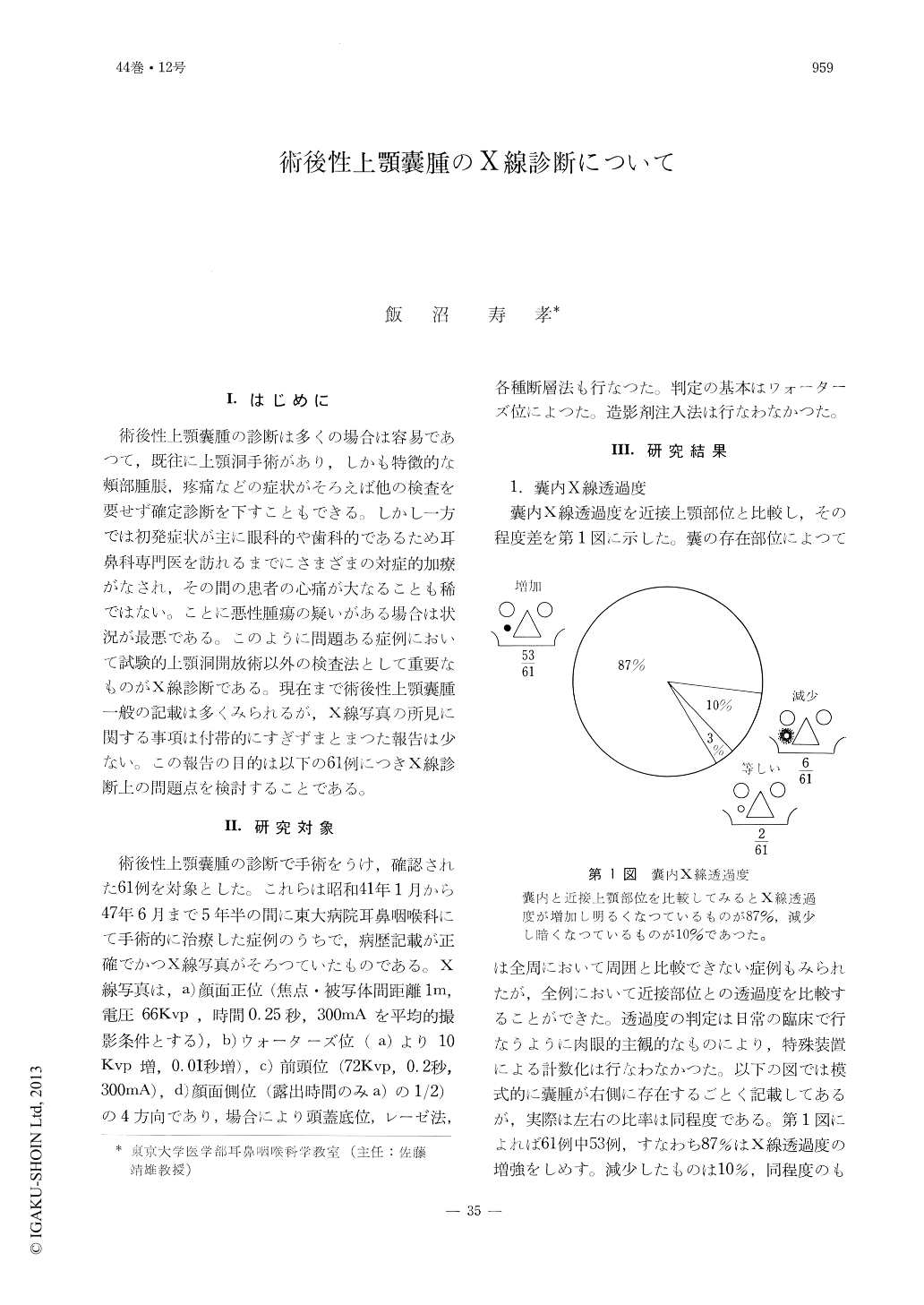
術後性上顎嚢腫の診断は多くの場合は容易であつて,既往に上顎洞手術があり,しかも特徴的な頬部腫脹,疼痛などの症状がそろえば他の検査を要せず確定診断を下すこともできる。しかし一方では初発症状が主に眼科的や歯科的であるため耳鼻科専門医を訪れるまでにさまざまの対症的加療がなされ,その間の患者の心痛が大なることも稀ではない。ことに悪性腫瘍の疑いがある場合は状況が最悪である。このように問題ある症例において試験的上顎洞開放術以外の検査法として重要なものがX線診断である。現在まで術後性上顎?腫一般の記載は多くみられるが,X線写真の所見に関する事項は付帯的にすぎずまとまつた報告は少ない。この報告の目的は以下の61例につきX線診断上の問題点を検討することである。
Sixty one cases of postoperative cysts of the maxilla are studied radiologically. Four routine films including PA, Water's, Caldwell's and lateral views were taken on each case. Water's view appeared to be the most informative for the present study. The characteristic x-ray findings were the increased radiolucency seen in 87% of cases and marginal sclerosis in 70%. Expansive changes of the cyst wall appear to be the main lesion that caused these x-ray findings. Contralateral to the affected or the operated side, cysts which were clinically silent were found in 34% of cases.
Copyright © 1972, Igaku-Shoin Ltd. All rights reserved.