Japanese
English
- 有料閲覧
PPV購入案内
1,320円 (1,200円+税10%) こちらは、573ページから580ページの文献です。購入には医書.jp本会員登録が必要です。
会員登録完了後に改めて上記「購入サイトへ」を選択してください。
または「購入サイトへ」選択後に「購入ログイン」、「新規会員登録」の順に進んでください。
会員登録について詳しくはをご確認ください。
- Abstract 文献概要
- 1ページ目 Look Inside
Ⅰ.はじめに
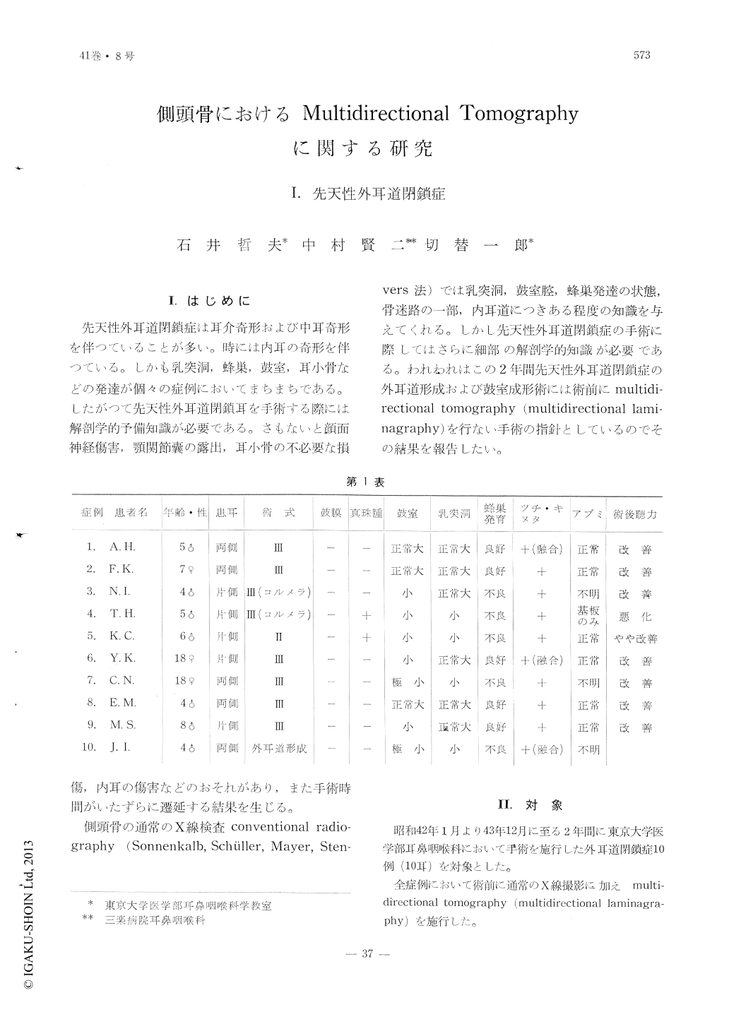
先天性外耳道閉鎖症は耳介奇形および中耳奇形を伴つていることが多い。時には内耳の奇形を伴つている。しかも乳突洞,蜂巣,鼓室,耳小骨などの発達が個々の症例においてまちまちである。したがつて先天性外耳道閉鎖耳を手術する際には解剖学的予備知識が必要である。さもないと顔面神経傷害,顎関節嚢の露出,耳小骨の不必要な損傷,内耳の傷害などのおそれがあり,また手術時間がいたずらに遷延する結果を生じる。
側頭骨の通常のX線検査conventional radiography(Sonnenkalb,Schuller,Mayer,Stenvers法)では乳突洞,鼓室腔,蜂巣発達の状態,骨迷路の一部,内耳道につきある程度の知識を与えてくれる。しかし先天性外耳道閉鎖症の手術に際してはさらに細部の解剖学的知識が必要である。われわれはこの2年間先天性外耳道閉鎖症の外耳道形成および鼓室成形術には術前にmultidirectional tomography(multidirectional laminagraphy)を行ない手術の指針としているのでその結果を報告したい。
Multidirectional tomography was carried out on 10 cases of congenital atresia of the external auditory meatus, in whom tympanoplastic surgery was performed on each. Tomography was done by Multiplanigraphy by Siemens by frontal plane with hypocycloidal movement.
Eight tomographic films were taken in 1mm thickness by 1mm interval. These pictures revealed the size of the tympanic cavity, the presence of the incus-malleus mass, the bony blockage of the atretic meatal wall, the oval window and the bony labyrinth.
In this way the preoperative tomographic findings were highly helpful in mapping out the plan of tympanoplasty upon these malformed ears.Type Ⅲ tympanoplasty was performed in 6 cases; type Ⅲ with columella in 2 eases and type Ⅱ in one case. An external auditory canal plasty alone was performed in one case.
Postoperative follow-up showed that 7 out of 10 cases revealed an improvement of hearing.
Copyright © 1969, Igaku-Shoin Ltd. All rights reserved.