Japanese
English
- 有料閲覧
PPV購入案内
1,320円 (1,200円+税10%) こちらは、649ページから655ページの文献です。購入には医書.jp本会員登録が必要です。
会員登録完了後に改めて上記「購入サイトへ」を選択してください。
または「購入サイトへ」選択後に「購入ログイン」、「新規会員登録」の順に進んでください。
会員登録について詳しくはをご確認ください。
- Abstract 文献概要
- 1ページ目 Look Inside
I.はじめに
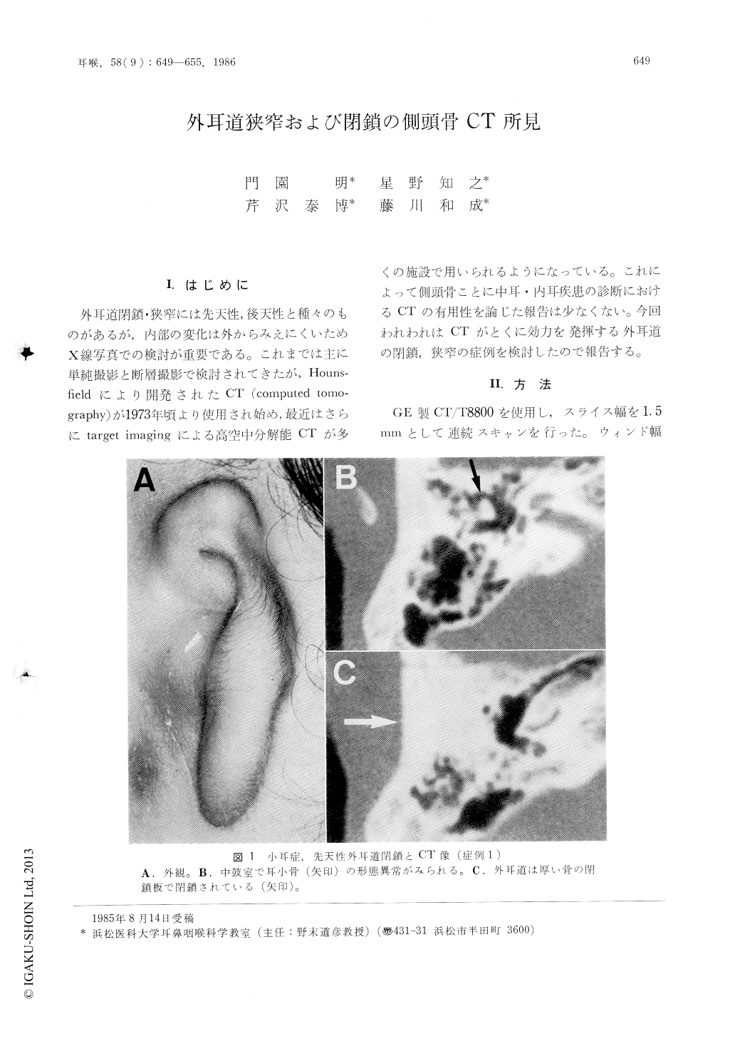
外耳道閉鎖・狭窄には先天性,後天性と種々のものがあるが,内部の変化は外からみえにくいためX線写真での検討が重要である。これまでは主に単純撮影と断層撮影で検討されてきたが,Hounsfieldにより開発されたCT(computed tomography)が1973年頃より使用され始め,最近はさらにtarget imagingによる高空中分解能CTが多くの施設で用いられるようになっている。これによって側頭骨ことに中耳・内耳疾患の診断におけるGTの有用性を論じた報告は少なくない。今回われわれはCTがとくに効力を発揮する外耳道の閉鎖,狭窄の症例を検討したので報告する。
CT findings of 5 cases of the stenotic external auditory canals were reported. Those cases included congenital atresia, carcinoma of the external and middle ear, malignant parotid tumor which invaded into external ear canal, traumatic disfigurement and protrusion of the condyle, and osteoma of the external auditory meatus. CT of the temporal bone was taken by a GE CT/T 8800 with targetimaging in 1.5mm horizontal sections. CT pictures clearly showed fine anatomical structures, extent of bone destruction and pathological disfigurement. CT was thought to give us the more valuable diagnostic informations than the conventional tomography in the cases with a narrowed external auditory canal.
Copyright © 1986, Igaku-Shoin Ltd. All rights reserved.