Japanese
English
- 有料閲覧
PPV購入案内
1,320円 (1,200円+税10%) こちらは、415ページから425ページの文献です。購入には医書.jp本会員登録が必要です。
会員登録完了後に改めて上記「購入サイトへ」を選択してください。
または「購入サイトへ」選択後に「購入ログイン」、「新規会員登録」の順に進んでください。
会員登録について詳しくはをご確認ください。
- Abstract 文献概要
- 1ページ目 Look Inside
I.緒言
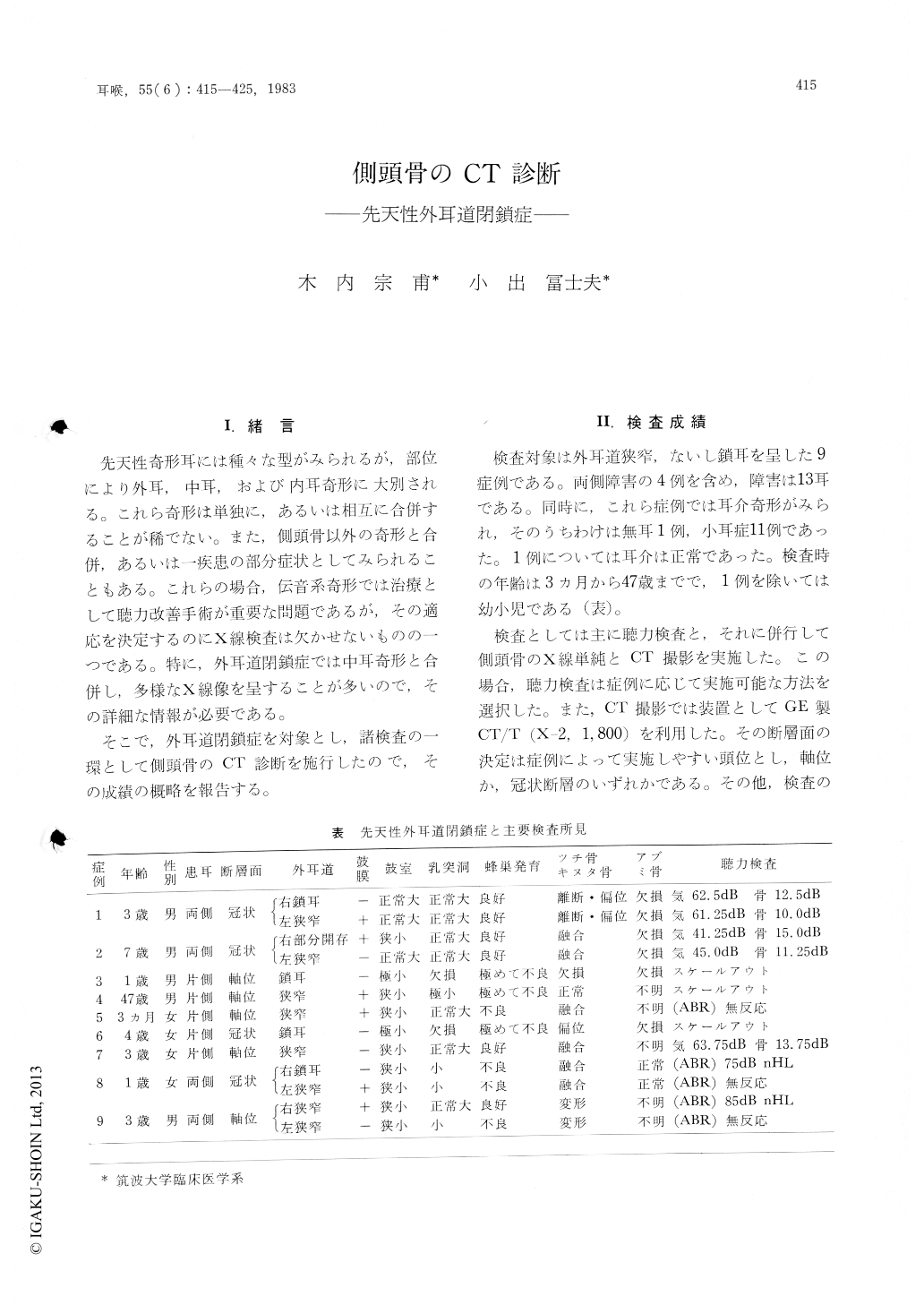
先天性奇形耳には種々な型がみられるが,部位により外耳,中耳,および内耳奇形に大別される。これら奇形は単独に,あるいは相互に合併することが稀でない。また,側頭骨以外の奇形と合併,あるいは一疾患の部分症状として入られることもある。これらの場合,伝音系奇形では治療として聴力改善手術が重要な問題であるが,その適応を決定するのにX線検査は欠かせないものの一つである。特に,外耳道閉鎖症では中耳奇形と合併し,多様なX線像を呈することが多いので,その詳細な情報が必要である。
そこで,外耳道閉鎖症を対象とし,諸検査の一環として側頭骨のCT診断を施行したので,その成績の概略を報告する。
Computed tomography was performed in 9 patients with atresia auris congenita. In this study, scans were done in axial or coronal projection with the CT/T body scanner.
The external auditory canals were involved in varying degrees from mild narrowing to complete atresia. In partial atresia, CT scanning demonstrated the patency of the canal, and also showed the eardrum as a clearly defined soft tissue. Simulatenously, the associated middle ear anomalies were well demonstrated. One of the most common deformities of the ossicular chains was fusion of the malleus and incus. In some cases, these ossicles were dislocated, and occasionally were fixed to the atresia plate or attic wall. Other variations of the temporal bone could be assumed.
We concluded that CT is the technique of choice to evaluate patients with atresia auris congenita.
Copyright © 1983, Igaku-Shoin Ltd. All rights reserved.