Japanese
English
- 有料閲覧
PPV購入案内
1,320円 (1,200円+税10%) こちらは、1015ページから1019ページの文献です。購入には医書.jp本会員登録が必要です。
会員登録完了後に改めて上記「購入サイトへ」を選択してください。
または「購入サイトへ」選択後に「購入ログイン」、「新規会員登録」の順に進んでください。
会員登録について詳しくはをご確認ください。
- Abstract 文献概要
- 1ページ目 Look Inside
I.はじめに
頭蓋骨内髄膜腫とはその発生部位あるいは主な腫瘍増殖部位が頭蓋骨内であるものを言い,1932年Atpers1)以降散発的な文献上の報告をみる.われわれの渉猟した限りわずか36例であった.術前のMRIが診断上有効であったと思われる頭蓋骨内髄膜腫の1例を経験したので若干の文献的考察を加え報告する.
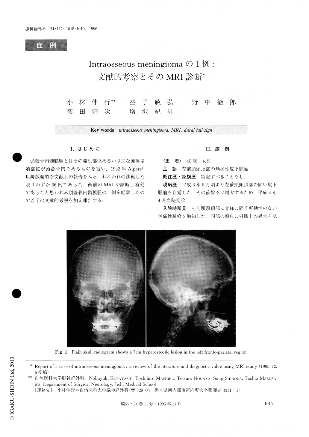
A 40-year-old woman with intraosseous meningioma is reported. She was admitted because of a slowly growing, bony hard, fixed mass in the temporal area that was covered by the normal scalp. The neurological examination on admission was negative. Plain skull radiogram demonstrated an area of hyperostotic change in the temporal region. Bone-targeted CT demonstrated irregularity in the structure of the skull bone. Selective external carotid angiogram showed a homogeneous tumor stain in the region of the skull thickening.
Macroscopically complete tumor resection (Simpson's grade 1) was carried out. Her postoperative course was uneventful. The skull tumor had the typical feature of meningotheliomatous meningioma. MRI showed a mass lesion of the temporal bone on T1-and T2-weighted im-ages, and a well-enhanced mass lesion of the outer dura and outer table of the skull bone on Gd-DTPA en-hanced T1 images. The findings of the preoperative MRI study using Gd-DTPA were identical with the tumor area on histological study. Therefore, MRI with Gd-DTPA enhancement was considered a useful di-agnostic modality.
Copyright © 1996, Igaku-Shoin Ltd. All rights reserved.