Japanese
English
- 有料閲覧
- Abstract 文献概要
- 1ページ目 Look Inside
- 参考文献 Reference
Ⅰ.はじめに
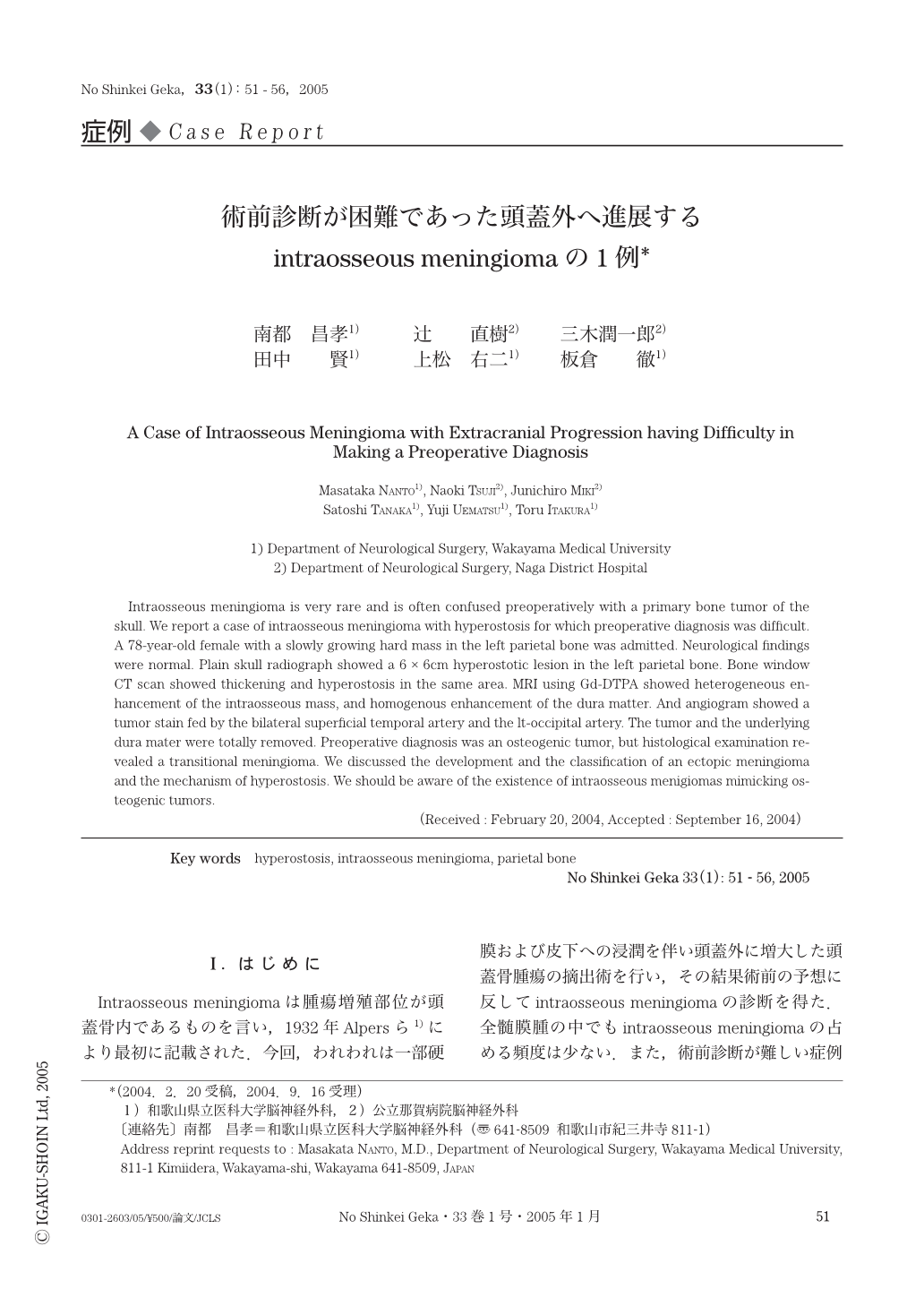
Intraosseous meningiomaは腫瘍増殖部位が頭蓋骨内であるものを言い,1932年Alpersら1)により最初に記載された.今回,われわれは一部硬膜および皮下への浸潤を伴い頭蓋外に増大した頭蓋骨腫瘍の摘出術を行い,その結果術前の予想に反してintraosseous meningiomaの診断を得た.全髄膜腫の中でもintraosseous meningiomaの占める頻度は少ない.また,術前診断が難しい症例が多いと考えられるため,文献的考察を加えて報告する.
Intraosseous meningioma is very rare and is often confused preoperatively with a primary bone tumor of the skull. We report a case of intraosseous meningioma with hyperostosis for which preoperative diagnosis was difficult. A 78-year-old female with a slowly growing hard mass in the left parietal bone was admitted. Neurological findings were normal. Plain skull radiograph showed a 6 × 6cm hyperostotic lesion in the left parietal bone. Bone window CT scan showed thickening and hyperostosis in the same area. MRI using Gd-DTPA showed heterogeneous enhancement of the intraosseous mass, and homogenous enhancement of the dura matter. And angiogram showed a tumor stain fed by the bilateral superficial temporal artery and the lt-occipital artery. The tumor and the underlying dura mater were totally removed. Preoperative diagnosis was an osteogenic tumor, but histological examination revealed a transitional meningioma. We discussed the development and the classification of an ectopic meningioma and the mechanism of hyperostosis. We should be aware of the existence of intraosseous menigiomas mimicking osteogenic tumors.

Copyright © 2005, Igaku-Shoin Ltd. All rights reserved.


