Japanese
English
- 有料閲覧
PPV購入案内
1,320円 (1,200円+税10%) こちらは、969ページから975ページの文献です。購入には医書.jp本会員登録が必要です。
会員登録完了後に改めて上記「購入サイトへ」を選択してください。
または「購入サイトへ」選択後に「購入ログイン」、「新規会員登録」の順に進んでください。
会員登録について詳しくはをご確認ください。
- Abstract 文献概要
- 1ページ目 Look Inside
- 参考文献 Reference
Ⅰ.はじめに
髄膜腫は原発性脳腫瘍の33.8%を占め15),多くが硬膜に沿って発生するが,ごく稀に硬膜とは無関係に骨原発に発生し,intraosseous meningioma(IM)として今までに複数の報告がある.原発性硬膜外髄膜腫の68%が頭蓋冠を含んでおり,IMに多いとされる部位は前頭頭頂部および眼窩部とされ3),術前の画像診断では,骨肉腫や線維性異形成,類骨骨腫との鑑別が問題となる.今回,術前画像検査で骨肉腫が疑われた頭蓋骨腫瘍が,病理診断でIMであった症例を経験したため,文献的考察を加え報告する.
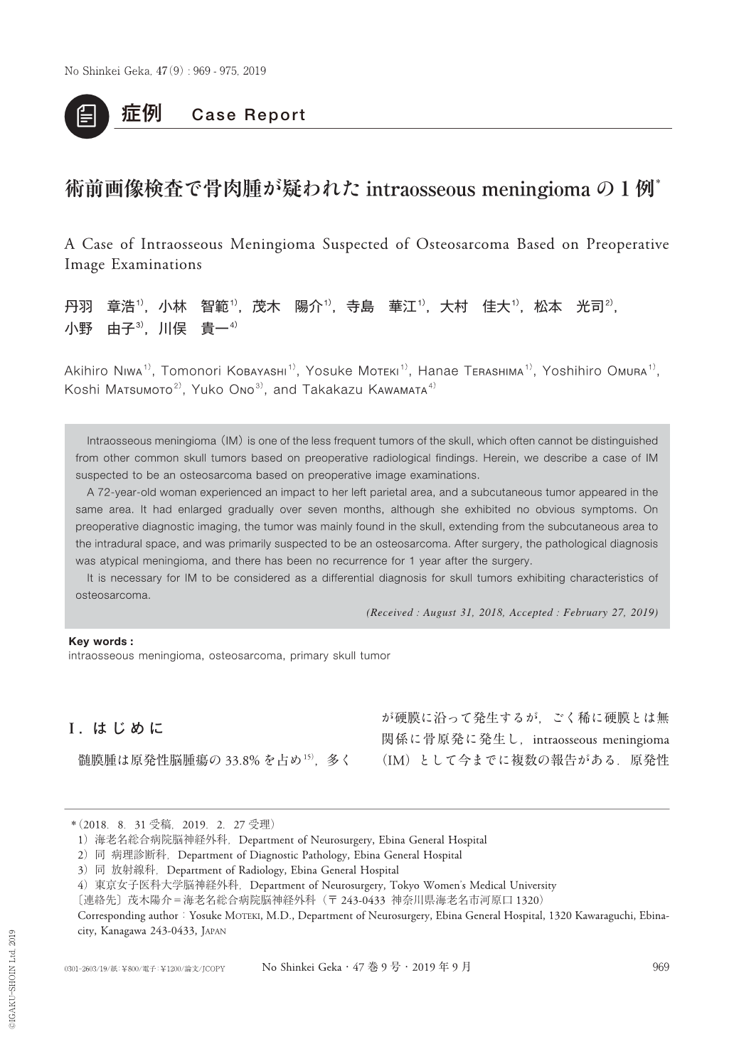
Intraosseous meningioma(IM)is one of the less frequent tumors of the skull, which often cannot be distinguished from other common skull tumors based on preoperative radiological findings. Herein, we describe a case of IM suspected to be an osteosarcoma based on preoperative image examinations.
A 72-year-old woman experienced an impact to her left parietal area, and a subcutaneous tumor appeared in the same area. It had enlarged gradually over seven months, although she exhibited no obvious symptoms. On preoperative diagnostic imaging, the tumor was mainly found in the skull, extending from the subcutaneous area to the intradural space, and was primarily suspected to be an osteosarcoma. After surgery, the pathological diagnosis was atypical meningioma, and there has been no recurrence for 1 year after the surgery.
It is necessary for IM to be considered as a differential diagnosis for skull tumors exhibiting characteristics of osteosarcoma.
Copyright © 2019, Igaku-Shoin Ltd. All rights reserved.