Japanese
English
- 有料閲覧
- Abstract 文献概要
- 1ページ目 Look Inside
I.はじめに
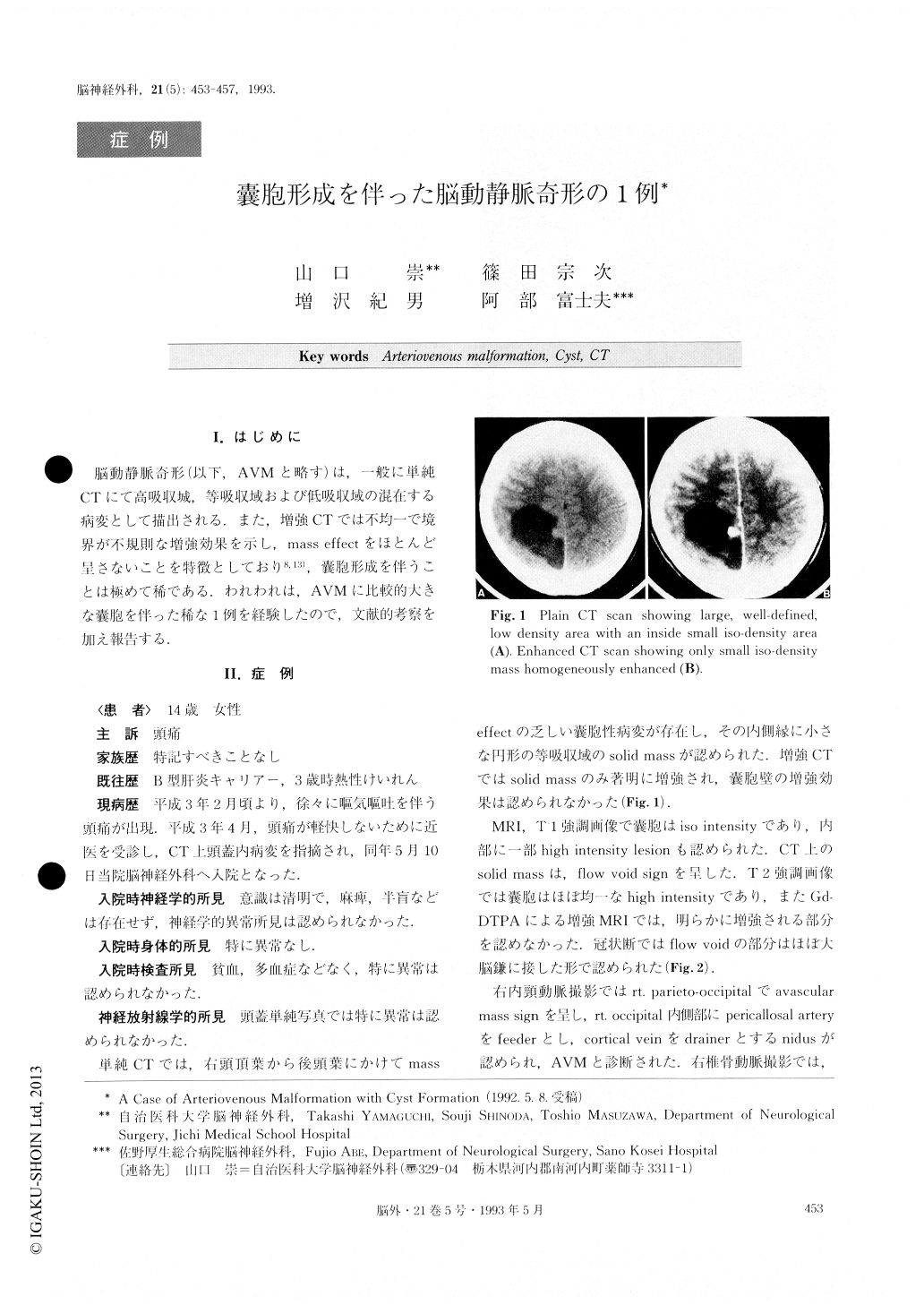
脳動静脈奇形(以下,AVMと略す)は,一般に単純CTにて高吸収城,等吸収域および低吸収域の混在する病変として描出される.また,増強CTでは不均一で境界が不規則な増強効果を示し,mass effectをほとんど呈さないことを特徴としており8,13),嚢胞形成を伴うことは極めて稀である.われわれは,AVMに比較的大きな嚢胞を伴った稀な1例を経験したので,文献的考察を加え報告する.
A case of arteriovenous malformation (AVM) with cyst formation is reported. A 14-year-old girl was admitted to our hospital, complaining of headache. On admission, CT scan showed a cystic mass with mural nodule in the right parietal lobe. Contrast enhanced CT scan demonstrated homogeneous enhancement of the nodule. MRI demonstrated the nodule as an area of signal void. Right carotid angiogram showed a typical nidus with feeding artery and draining vein. The nidus was totally removed by the trans-cystic approach. Histological diagnosis was typical AVM with hemosiderin deposits.
AVM with cyst formation is very rare, with only six cases having been reported. The mechanism of cyst formation is controversial. Past reports have hypothesized the participation of a massive hemorrhage, or exudative process. Our case showed hemosiderin deposits, but no symptom indicative of hemorrhage. In this case, the etiology of cyst formation was considered to be exudation and thrombosis of the nidus.

Copyright © 1993, Igaku-Shoin Ltd. All rights reserved.


