Japanese
English
症例
Intraosseous meningiomaの1例
Report of A Case of Intraosseous Meningioma
金子 文仁
1,4
,
高瀬 憲作
1
,
西山 一英
1
,
日下 和昌
1
,
森住 啓
2
,
松本 圭蔵
3
Fumihito KANEKO
1,4
,
Kensaku TAKASE
1
,
Kazuhide NISHIYAMA
1
,
Kazumasa KUSAKA
1
,
Hiroshi MORIZUMI
2
,
Keizo MATSUMOTO
3
1徳島市民病院脳神経外科
2徳島市民病院病理
3徳島大学脳神経外科
4現籍 中村市立市民病院脳神経外科
1Department of Neurosurgery, Tokushima Municipal Hospital
2Department of Pathology, Tokushima Municipal Hospital
3Department of Neurological Surgery, School of Medicine, The University of Tokushima
キーワード:
Intraosseous meningioma
,
Skull tumor
,
Histogenesis
Keyword:
Intraosseous meningioma
,
Skull tumor
,
Histogenesis
pp.197-202
発行日 1988年2月10日
Published Date 1988/2/10
DOI https://doi.org/10.11477/mf.1436202548
- 有料閲覧
- Abstract 文献概要
- 1ページ目 Look Inside
I.はじめに
頭蓋骨に主座を置くmeningiomaは,intra—osseous meningiomaとも呼ばれ2,3,7,11,13,17,19),その報告は1932年Alpersら1)が最初に記載して以来散見される.今回われわれは,intra—osseous meningiomaと思われる1例を経験したので,若干の文献的考察を加えて報告する.
Cases of intraosseous meningioma appear to be very rare. In the present paper, we report such a case and discuss its etiological histogenesis on the basis of a re-view of 26 cases previously reported.
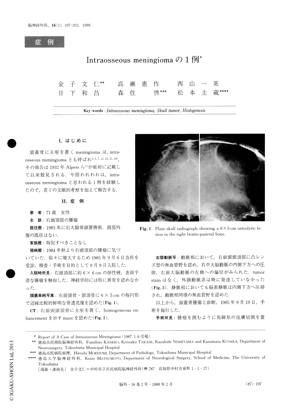
A 71-year-old famale was admitted to our department because of a painless mass in the right parietal region. Neurological findings were normal. Plain skull radio-graph showed a 6×5-cm osteolytic lesion in the right fronto-parietal bone. CT scan demonstrated this lesion as a mass showing homogeneous enhancement with contrast medium.

Copyright © 1988, Igaku-Shoin Ltd. All rights reserved.


