Japanese
English
- 有料閲覧
PPV購入案内
1,320円 (1,200円+税10%) こちらは、55ページから61ページの文献です。購入には医書.jp本会員登録が必要です。
会員登録完了後に改めて上記「購入サイトへ」を選択してください。
または「購入サイトへ」選択後に「購入ログイン」、「新規会員登録」の順に進んでください。
会員登録について詳しくはをご確認ください。
- Abstract 文献概要
- 1ページ目 Look Inside
I.はじめに
定位脳手術は1947年Spiegd & Wycisら28)により初めて施行され,最小の外科的侵襲により目標部位への到達が可能な方法として発展してきた.さらに1972年,CTが初めて臨床例に用いられたが1),その後CTを定位脳手術に応用する研究が進められ,脳腫瘍,脳内出血,脳膿瘍など,CTによって病巣部の正確な拡がりを明らかにできるようになった疾患に対する定位脳手術が容易になった.
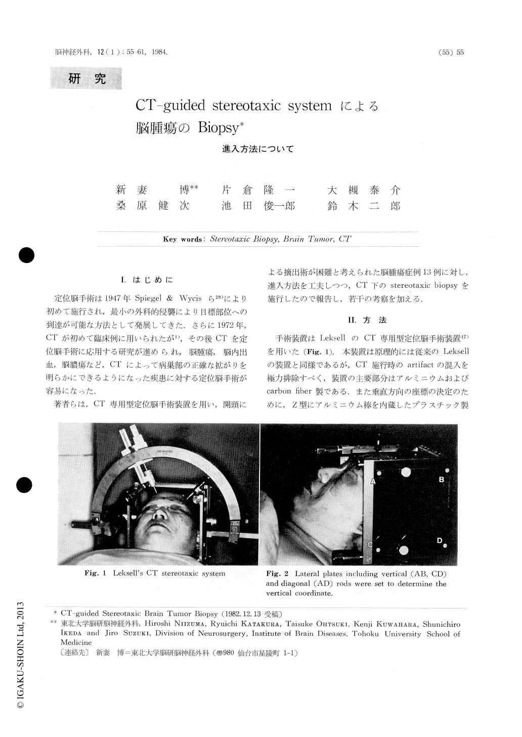
著者らは,CT専用型定位脳手術装置を用い,開頭による摘出術が困難と考えられた脳腫瘍症例13例に対し,進入方法を工夫しつつ,CT下のstereotaxic biopsyを施行したので報告し,若干の考察を加える.
CT-guided stereotaxic biopsy was performed on 13cases of the brain tumor using Leksell's CT stereotaxicsystem under control of Hitachi W-3 scanner.
Trepanation was put on near to the coronal suture,and biopsy needle was inserted parallel to sagittalplane in two cases. The course of the needle wasshowed by the sagittal reconstructive CT. In 11cases, biopsy needle was inserted parallel to the CTslice. Trepanation was put on frontal area in ninecases of frontal tumor, and on posterior temporal areain four cases of occipital or thalamic tumor. Thecourse of needle was showed on one CT slice.
Copyright © 1984, Igaku-Shoin Ltd. All rights reserved.