Japanese
English
- 有料閲覧
- Abstract 文献概要
- 1ページ目 Look Inside
Ⅰ.はじめに
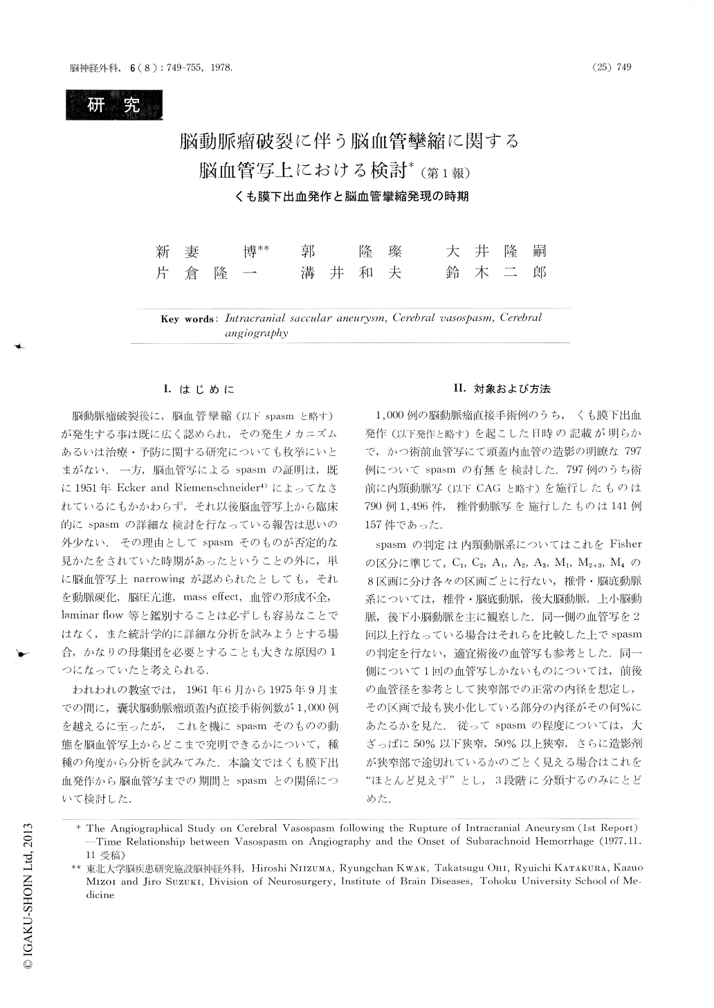
脳動脈瘤破裂後に,脳血管攣縮(以下spasmと略す)が発生する事は既に広く認められ,その発生メカニズムあるいは治療・予防に関する研究についても枚挙にいとまがない.一方,脳血管写によるspasmの証明は,既に1951年Ecker and Riemenschneider4)によってなされているにもかかわらず,それ以後脳血管写上から臨床的にspasmの詳細な検討を行なっている報告は思いの外少ない.その理由としてSpasmそのものが否定的な見かたをされていた時期があったということの外に,単に脳血管写上narrowingが認められたとしても,それを動脈硬化,脳圧亢進,mass effect,血管の形成不全,laminar fiow等と鑑別することは必ずしも容易なことではなく,また統計学的に詳細な分析を試みようとする場合,かなりの母集団を必要とすることも大きな原因の1つになっていたと考えられる.
われわれの教室では,1961年6月から1975年9月までの間に,嚢状脳動脈瘤頭蓋内直接手術例数が1,000例を越えるに至ったが,これを機にSpasmそのものの動態を脳血管写上からどこまで究明できるかについて,種種の角度から分析を試みてみた.本論文ではくも膜下出血発作から脳血管写までの期間とspasmとの関係について検討した.
Time relationship between the presence of vasospasm on angiography and the onset of suharachnoid hemorrhage (SAH) was investigated with 797 direct surgical cases with intracranial saccular aneurysm.
It has been said that vasospasm would occur in early days after the onset of SAH. But in this paper, in the cases with only one history of SAH, vasospasm occurred within 3 days after SAH was seen in only 4.2% of the 120 cases. The peak of vasospasm on angiography was seen in the period between 10 and 17 days after SAH. During this period vasospasm was seen in 49.1% of the 116 cases.

Copyright © 1978, Igaku-Shoin Ltd. All rights reserved.


