Japanese
English
- 有料閲覧
PPV購入案内
1,320円 (1,200円+税10%) こちらは、1017ページから1022ページの文献です。購入には医書.jp本会員登録が必要です。
会員登録完了後に改めて上記「購入サイトへ」を選択してください。
または「購入サイトへ」選択後に「購入ログイン」、「新規会員登録」の順に進んでください。
会員登録について詳しくはをご確認ください。
- Abstract 文献概要
- 1ページ目 Look Inside
I.はじめに
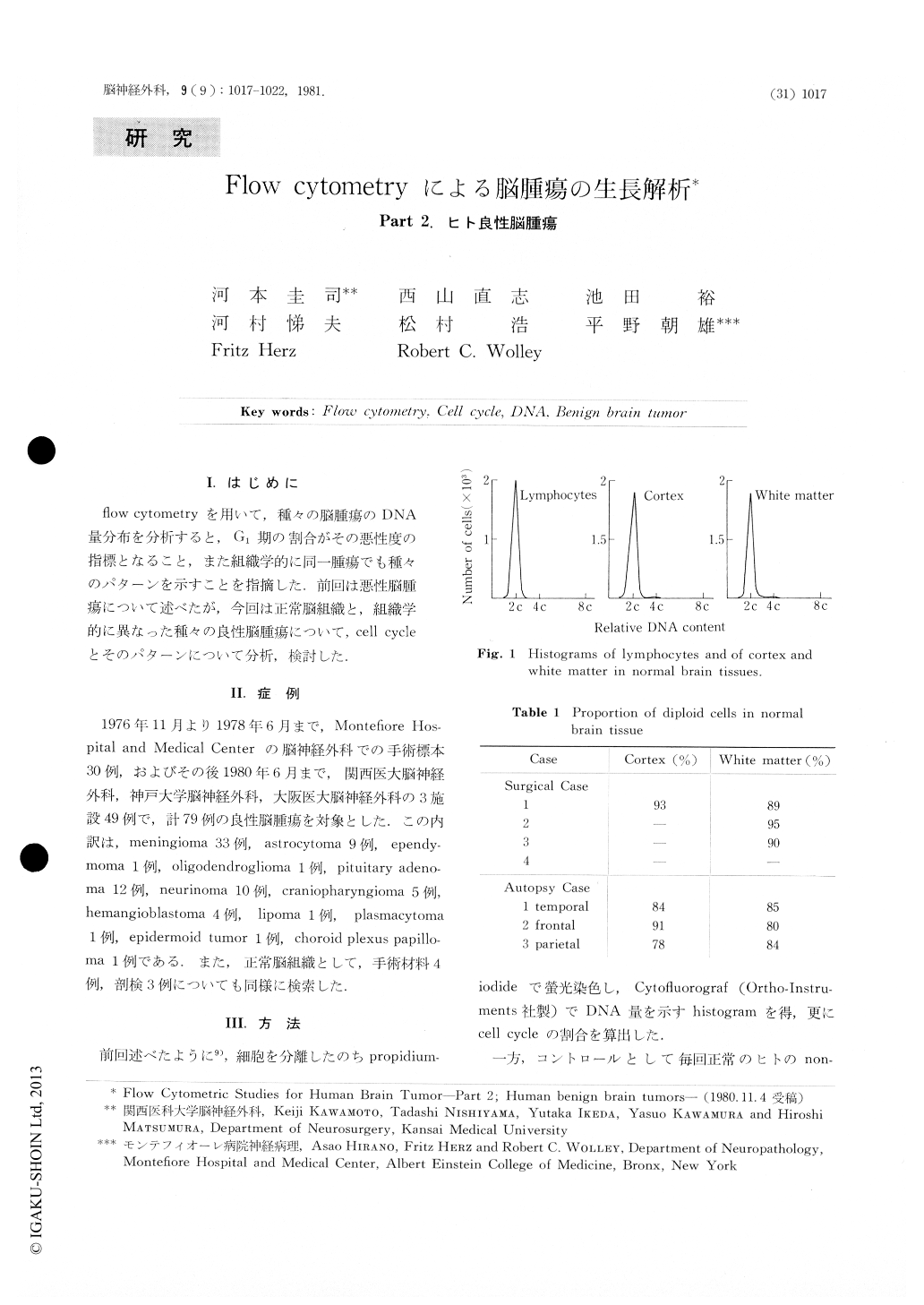
fiow cytometryを用いて,種々の脳腫瘍のDNA量分布を分析すると,G1期の割合がその悪性度の指標となること,また組織学的に同一腫瘍でも種々のパターンを示すことを指摘した.前回は悪性脳腫瘍について述べたが,今回は正常脳組織と,組織学的に異なった種々の良性脳腫瘍について,cell cycleとそのパターンについて分析,検討した.
This report compares the DNA content distribution of normal human brain tissue and of benign tumors of the brain. The cells were obtained from seven normal specimens and from fourteen types of benign brain tumors of 79 patients. The DNA content was determined by flow cytometry on single cell suspensions. Propidium iodide was used as DNA-intercalating fluorochrome. Normal, non-stimulated lymphocytes served as diploid controls.
The proportions of diploid cells in G1 ranged from 78% to 93% and from 80% to 95% in the normal cortex and normal white matter, respectively. In the specimens of benign brain tumor the range was from 70% to 98%.
Copyright © 1981, Igaku-Shoin Ltd. All rights reserved.