Japanese
English
- 有料閲覧
PPV購入案内
1,320円 (1,200円+税10%) こちらは、645ページから648ページの文献です。購入には医書.jp本会員登録が必要です。
会員登録完了後に改めて上記「購入サイトへ」を選択してください。
または「購入サイトへ」選択後に「購入ログイン」、「新規会員登録」の順に進んでください。
会員登録について詳しくはをご確認ください。
- Abstract 文献概要
- 1ページ目 Look Inside
Ⅰ.はじめに
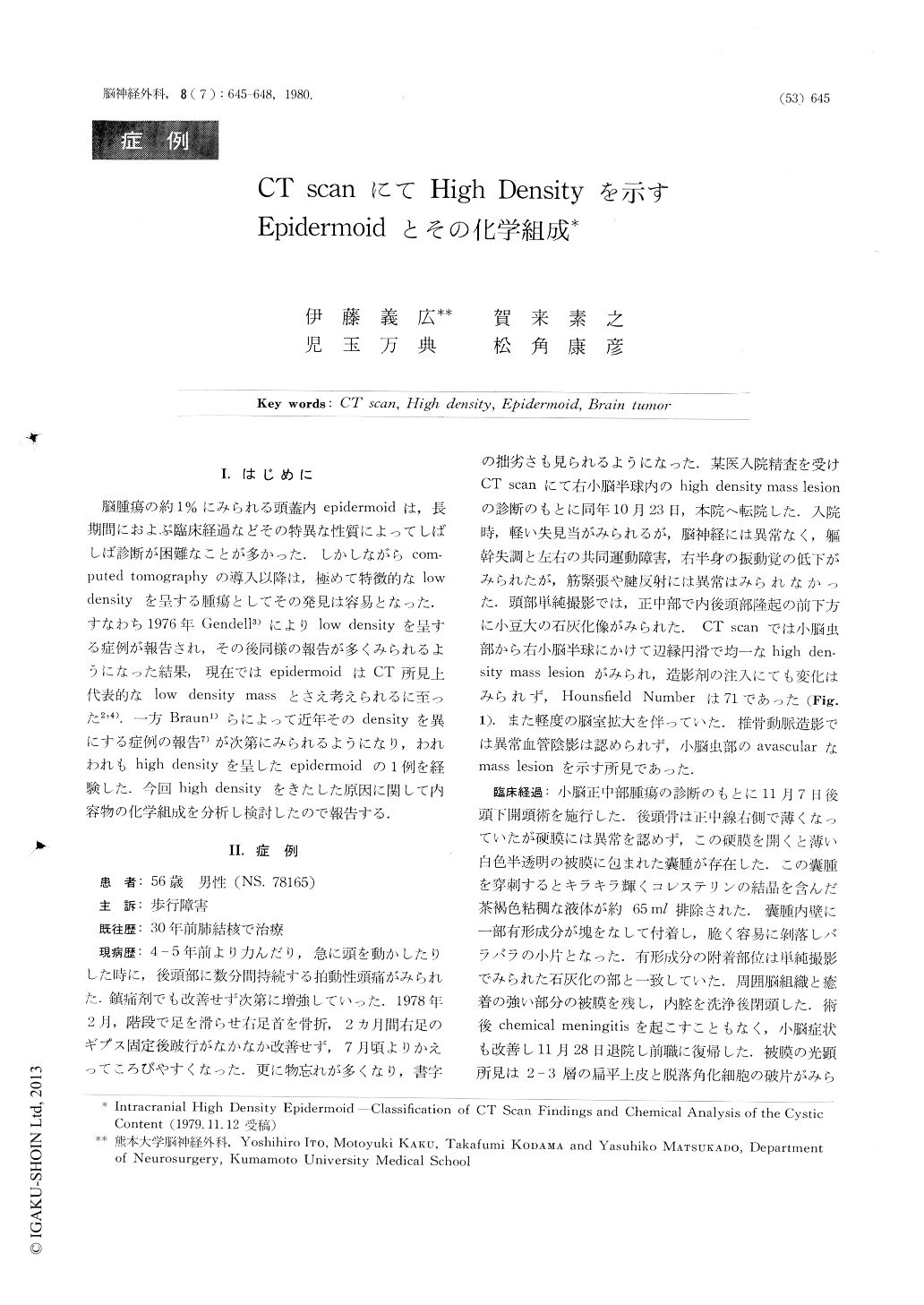
脳腫瘍の約1%にみられる頭蓋内epidermoidは,長期間におよぶ臨床経過などその特異な性質によってしばしば診断が困難なことが多かった.しかしながらcomputed tomographyの導入以降は,極めて特徴的なlowdensityを呈する腫瘍としてその発見は容易となった.すなわち1976年Gendell3)によりlow densityを呈する症例が報告され,その後同様の報告が多くみられるようになった結果,現在ではepidermoidはCT所見上代表的なlow density massとさえ考えられるに至った2,4).一方Braun1)らによって近年そのdensityを異にする症例の報告7)が次第にみられるようになり,われわれもhigh densityを呈したepidermoidの1例を経験した,今回high densityをきたした原因に関して内容物の化学組成を分析し検討したので報告する.
The author described an epidermoid which revealed homogenous high density in CT scan without contrast enhancement. Sixteen reported cases of intracranial epidermoid with CT scan including our 4 cases were classified in three types; Type classical pearly tumor of low density with no contrast enhancement. Type II; low density mass with marginal high density ring by contrast enhancement. Type III; homogenous high density mass with no contrast enhancement. The reason of high density in CT scan was considered mainly clue to the peculiar cystic content, which was brown in color and gelatinous.
Copyright © 1980, Igaku-Shoin Ltd. All rights reserved.