Japanese
English
- 有料閲覧
PPV購入案内
1,320円 (1,200円+税10%) こちらは、333ページから341ページの文献です。購入には医書.jp本会員登録が必要です。
会員登録完了後に改めて上記「購入サイトへ」を選択してください。
または「購入サイトへ」選択後に「購入ログイン」、「新規会員登録」の順に進んでください。
会員登録について詳しくはをご確認ください。
- Abstract 文献概要
- 1ページ目 Look Inside
Ⅰ.緒言
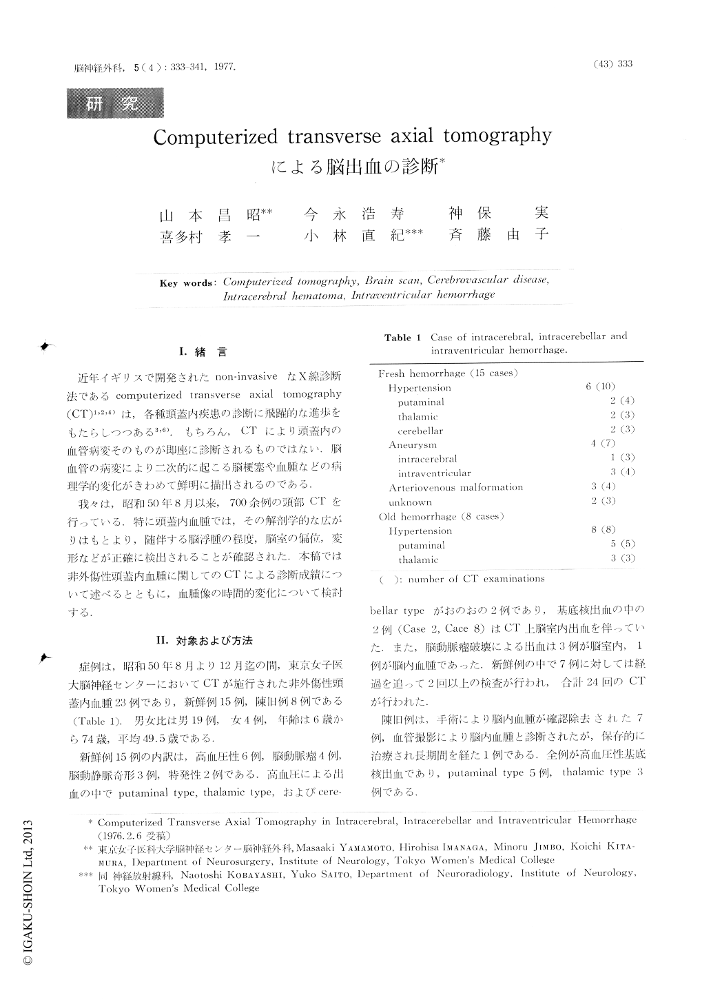
近年イギリスで開発されたnon-invasiveなX線診断法であるcomputerized transverse axial tomography(CT)1,2,4)は,各種頭蓋内疾患の診断に飛躍的な進歩をもたらしつつある3,6).もちろん,CTにより頭蓋内の血管病変そのものが即座に診断されるものではない.脳血管の病変により二次的に起こる脳梗塞や血腫などの病理学的変化がきわめて鮮明に描出されるのである.
我々は,昭和50年8月以来,700余例の頭部CTを行っている.特に頭蓋内血腫では,その解剖学的な広がりはもとより,随伴する脳浮腫の程度,脳室の偏位,変形などが正確に検出されることが確認された.本稿では非外傷性頭蓋内血腫に関してのCTによる診断成績について述べるとともに,血腫像の時間的変化について検討する.
Computerized transverse axial tomography (CT) of the brain is a recently developed method which allows noninvasive roentgenologic evaluation of intracranial diseases. The advent of CT represents a great advance in the diagnosis of a very wide variety of intracranial lesions, including cerebrovascular diseases. Especially, CT was found to be extremely informative in evaluating intracerebral, intracerebellar and intraventricular hemorrhage.
The purpose of this report is to evaluate the clinical usefulness of CT in the diagnosis of intracramal hemor rhage.
Copyright © 1977, Igaku-Shoin Ltd. All rights reserved.