Japanese
English
- 有料閲覧
- Abstract 文献概要
- 1ページ目 Look Inside
気脳術にさいしcoronalおよびsagittal tomographyに加えてaxial transverse tomographyを25例につき施行し,本法がcoronalおよびsaglttal tomographyよりもすぐれている点を求めた。
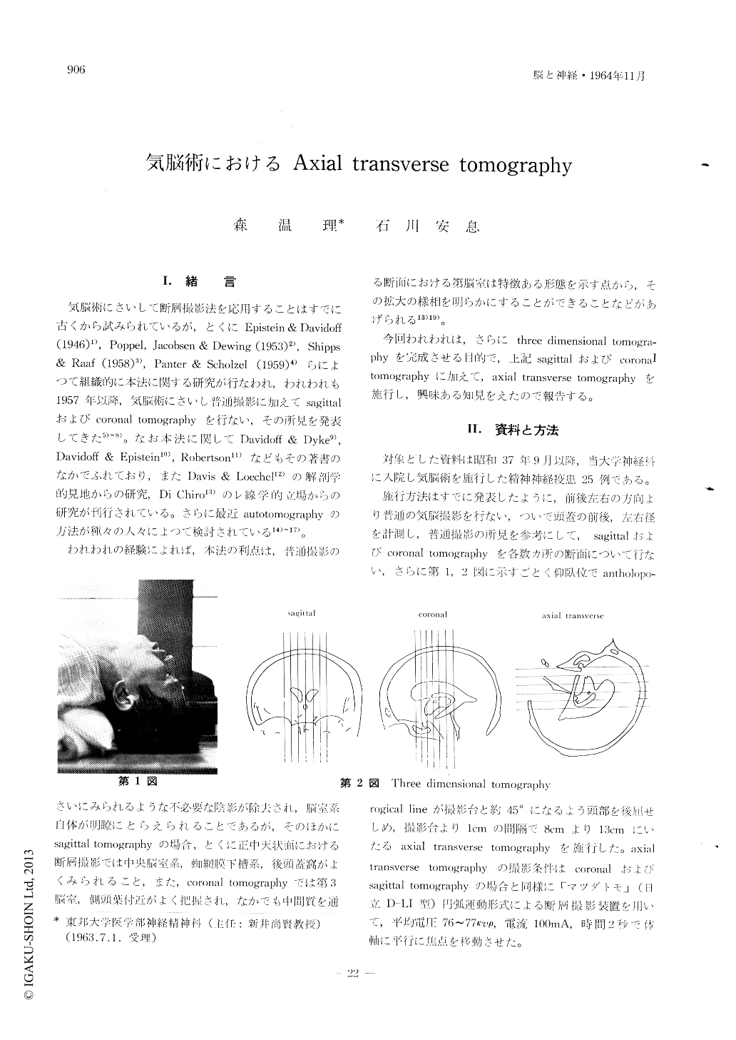
撮影方法はcoronalおよびsagittal tomography施行後,仰臥位にてalltholoporogical lineが撮影台と45°になるよう頭部を後方に曲げ,1cmおきに撮影台より3cmより13cmにいたるaxial transverse tomographyを行なつた。
その結果coronalおよびsagittal tomographyよりも両側同時に広範囲の脳室系および頭蓋底の重要な部分を把握できることが明らかとなり,これにより病変の診断に役だちうるものと考えられた。
Coronal and sagittal laminagraphy in PEG has been used for several years in our clinic. In order to complete the laminagraphic method, however, axial transverse laminagraphy is also necessary; because the coronal, sagittal and transverse planes are three dimensions of the head.
The patient was lied supineely and his head was placed in hyperextension and the angle between anthropological plane and film was 45. This halfa-xial position of the head was practicable for all patients. The laminagram was taken per 1 cm through the planes of 8 to 13 cm from the table top.
The main visualized brain regions by this method were lateral ventricles, third and fourth ventricles, aqueduct, cisterna magna, fissura cerebri lateralis, insular sulcus, cisterna chiasmatis, cisterna inter-peduncularis, vallecula cerebri lateralis and skull basis. It was worthy of notice that the brain regions around skull basis was more clearly visualized by this method than by other methods, namely coronal and sagittal methods. In this report, the films of axial transverge laminagraphy in twenty five cases were analysed and the usefulness of this method was discussed.

Copyright © 1964, Igaku-Shoin Ltd. All rights reserved.


