Japanese
English
- 有料閲覧
PPV購入案内
1,320円 (1,200円+税10%) こちらは、623ページから631ページの文献です。購入には医書.jp本会員登録が必要です。
会員登録完了後に改めて上記「購入サイトへ」を選択してください。
または「購入サイトへ」選択後に「購入ログイン」、「新規会員登録」の順に進んでください。
会員登録について詳しくはをご確認ください。
- Abstract 文献概要
- 1ページ目 Look Inside
Ⅰ.はじめに
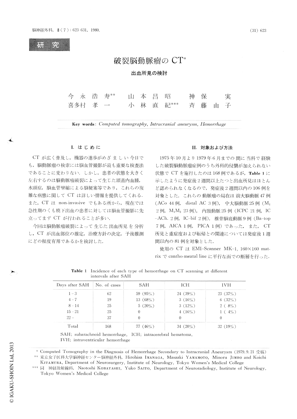
CTが広く普及し,機器の進歩がめざましい今日でも,脳動脈瘤の検索には脳血管撮影が最も重要な検査法であることに変わりない.しかし,患者の状態を大きく左右するのは脳動脈瘤破裂によって生じた頭蓋内血腫,水頭症,脳血管攣縮による脳梗塞等であり,これらの複雑な病態に関してCTは詳しい情報を提供してくれる.また,CTはnon-invasiveでもある所から,現在では急性期のくも膜下出血の患者に対しては脳血管撮影に先立ってまずCTが行われることが多い.
今回は脳動脈瘤破裂によって生じた出血所見を分析し,CTが出血.部位の推定,治療方針の決定,予後推測にどの程度有用であるかを検討した.
In 168 patients with ruptured intracranial aneurysms, the pathology of intracranial hemorrhage visualized on CT was analyzed.
Blood in the subarachnoid space could be visualized in 95% of cases within three days after SAH and 75% of 106 cases within two weeks after SAH. In one case blood clot in the subarachnoid space visible up to 13 days after SAH. Concerning the cases within two weeks after the bleeding, intracerebral hematomas were observed in 36% of anterior cerebral aneurysms and middle cerebral aneurysms, 16% of internal carotid aneurysms and none of vertebro-basilar aneurysms.
Copyright © 1980, Igaku-Shoin Ltd. All rights reserved.