Japanese
English
- 有料閲覧
PPV購入案内
1,320円 (1,200円+税10%) こちらは、497ページから510ページの文献です。購入には医書.jp本会員登録が必要です。
会員登録完了後に改めて上記「購入サイトへ」を選択してください。
または「購入サイトへ」選択後に「購入ログイン」、「新規会員登録」の順に進んでください。
会員登録について詳しくはをご確認ください。
- Abstract 文献概要
- 1ページ目 Look Inside
Ⅰ.はじめに
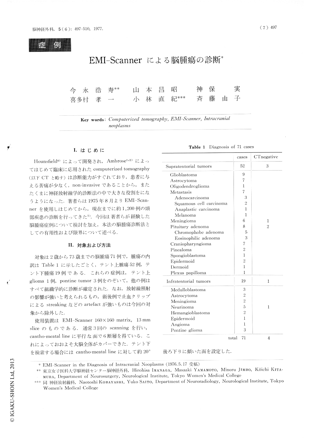
Hounsfield4)によって開発され,Ambrose1,2)によってはじめて臨床に応用されたcomputerized tomography(以下CTと略す)は診断能力がすぐれており,患者に与える苦痛が少なく,non-invasiveであることから,またたくまに神経放射線学的診断法の中で大きな役割をになうようになった.著者らは1975年8月よりEMI-Scannerを使用しはじめてから,現在までに約1,200例の頭部疾患の診断を行ってきた7).今回は著者らが経験した脳腫瘍症例について検討を加え,本法の脳腫瘍診断法としての有用性および限界について述べる.
The diagnostic results of EMI-Scanner in 71 cases of intracranial neoplasms were presented. In 67 cases localization and extensions of the tumor were correctly diagnosed with the EMI-Scanner. In four false negative cases, one case of vertex meningioma extending mainly out of the cranium, two cases of intrasellar pituitary adenoma and one case of acoustic neurinoma of 1.5 cm in diameter were included.
The characteristic features of the various tumors were summerized as follows:
Copyright © 1977, Igaku-Shoin Ltd. All rights reserved.