Japanese
English
- 有料閲覧
- Abstract 文献概要
- 1ページ目 Look Inside
Ⅰ.はじめに
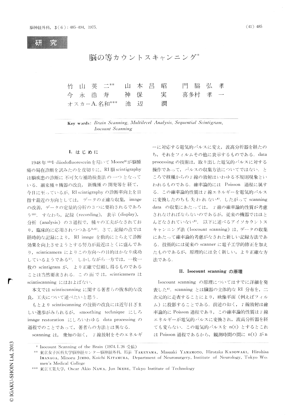
1948年131I-diiodofluoresceinを用いてMoore8)が脳腫瘍の局在診断を試みたのを皮切りに,Rl脳scintigraphyは脳疾患の診断に不可欠な補助検査法の一つとなっている.爾来種々機器の改良,新機種の開発等を経て,今日に至っているが,Rl scintigraphyの診断率向上を目指す最近の方向としては,データの正確な収集,imageの改善,デーケの定量的分祈の3つに要約されるであろう10).すなわち記録(recording),表示(display),分析(analysis)の3過程で,種々の工夫がなされており,臨床的に応用されつつある3,6).さて,記録の点では経時的な記録により,RI mageを動的にとらえて診断効果を向上させようとする努力が最近はとくに盛んであり,scinticameraによりこの方向への目的はかなり成功しているようである1)しかしながら一方では,一枚一枚のscintigramが,より正確で信頼し得るものであることは当然要求される.この面では,scinticameraはscintiscanningにはおよばない.
本文ではscintiscanningに関する著者らの抜本的な改良,工夫について述べたいと思う.
It is well known that the gamma ray emitted by a radionuclide is a Poisson process, so its statistic nature should be taken in consideration when the scintiscanning is performed. But very few attempts have been clone from this point of view.
After examining in detail the imaging pulsecharactristics, the authors introduced a mean for evaluating the fidelity of the collected data, and based on this criterion, pointed out some problems in the conventional scanning method and finally described a statistical way of improving the scintigram.

Copyright © 1975, Igaku-Shoin Ltd. All rights reserved.


