Japanese
English
研究
大脳半球腫瘍CTのHistogram解析
Histographic Analysis of Computed Tomography in Cerebral Hemispheric Tumors
今永 浩寿
1
,
山本 昌昭
1
,
神保 実
1
,
小林 直紀
2
,
斉藤 由子
2
,
喜多村 孝一
3
Hirohisa IMANAGA
1
,
Masaaki YAMAMOTO
1
,
Minoru JIMBO
1
,
Naotoshi KOBAYASHI
2
,
Yuko SAITO
2
,
Koiti KITAMURA
3
1東京女子医科大学第二病院脳神経外科
2東京女子医科大学脳神経センター神経放射線科
3東京女子医科大学脳神経センター脳神経外科
1Department of Neurosurgery, Ogu Hospital, Tokyo Women's Medical College
2Department of Neuroradiology, Institute of Neurology, Tokyo Women's Medical College
3Department of Neurosurgery, Institute of Neurology Tokyo Women's Medical College
キーワード:
Computed tomography
,
Cerebra
,
tumor
,
Histogram
Keyword:
Computed tomography
,
Cerebra
,
tumor
,
Histogram
pp.481-488
発行日 1983年5月10日
Published Date 1983/5/10
DOI https://doi.org/10.11477/mf.1436201669
- 有料閲覧
- Abstract 文献概要
- 1ページ目 Look Inside
I.はじめに
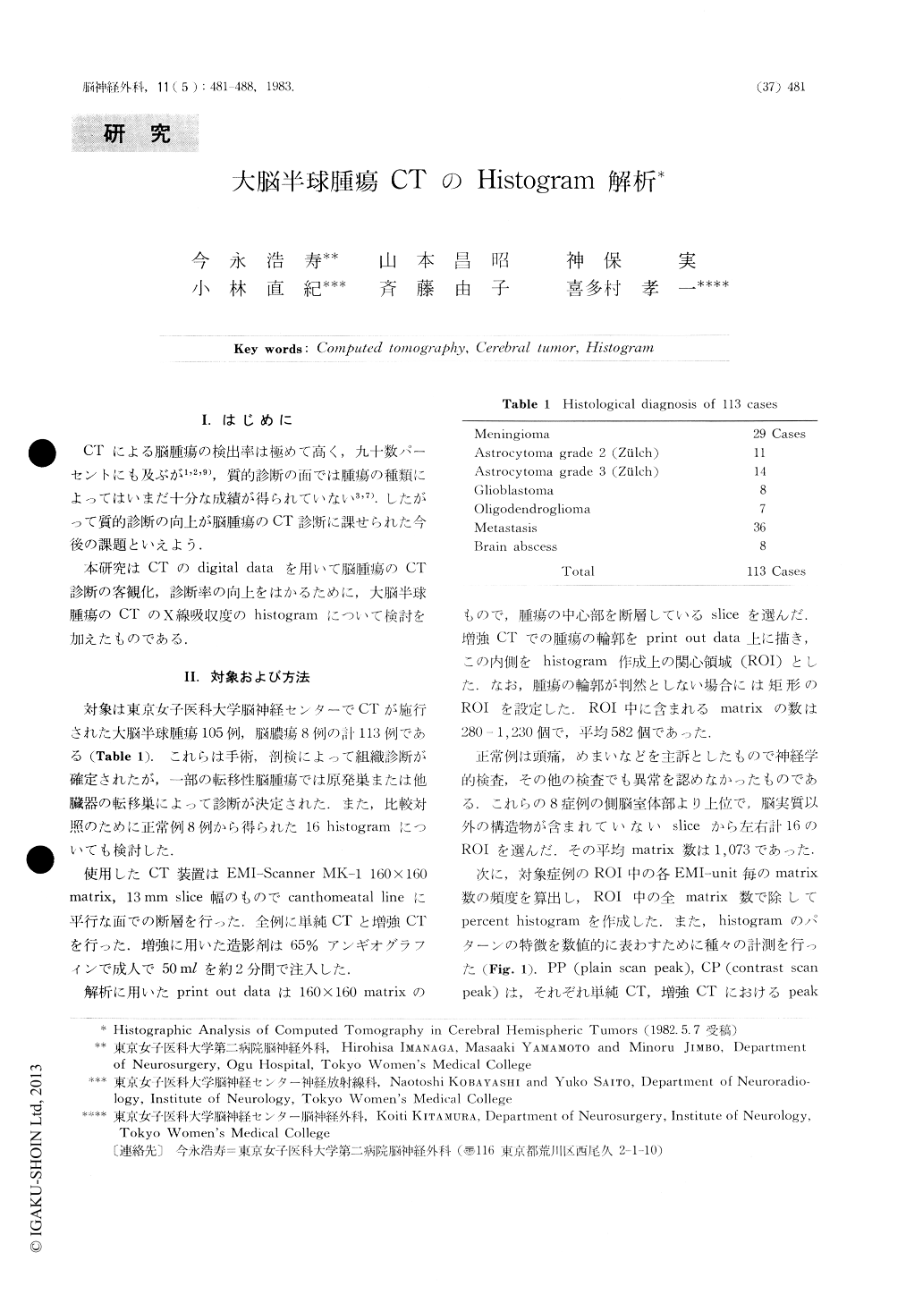
CTによる脳腫瘍の検出率は極めて高く,九十数パーセントにも及ぶが1,2,9),質的診断の面では腫瘍の種類によってはいまだ十分な成績が得られていない3,7).したがって質的診断の向上が脳腫瘍のCT診断に課せられた今後の課題といえよう.
本研究はCTのdigital dataを用いて脳腫瘍のCT診断の客観化,診断率の向上をはかるために,大脳半球腫瘍のCTのX線吸収度のhistogramについて検討を加えたものである.
The aim of this report is to assess the possibilityof digital processing of computed tomography forhistological diagnosis of brain tumors.
For this purpose, percent histograms of x-rayattenuation value of lesions were analysed in 113 casesof histologically verified hemispheric tumors. Sixteenhistograms obtained from normal brain parenchymaserved as controls. Histograms were made from thesame slices before and after contrast enhancement.

Copyright © 1983, Igaku-Shoin Ltd. All rights reserved.


