Japanese
English
- 有料閲覧
PPV購入案内
1,320円 (1,200円+税10%) こちらは、497ページから510ページの文献です。購入には医書.jp本会員登録が必要です。
会員登録完了後に改めて上記「購入サイトへ」を選択してください。
または「購入サイトへ」選択後に「購入ログイン」、「新規会員登録」の順に進んでください。
会員登録について詳しくはをご確認ください。
- Abstract 文献概要
- 1ページ目 Look Inside
I.はじめに
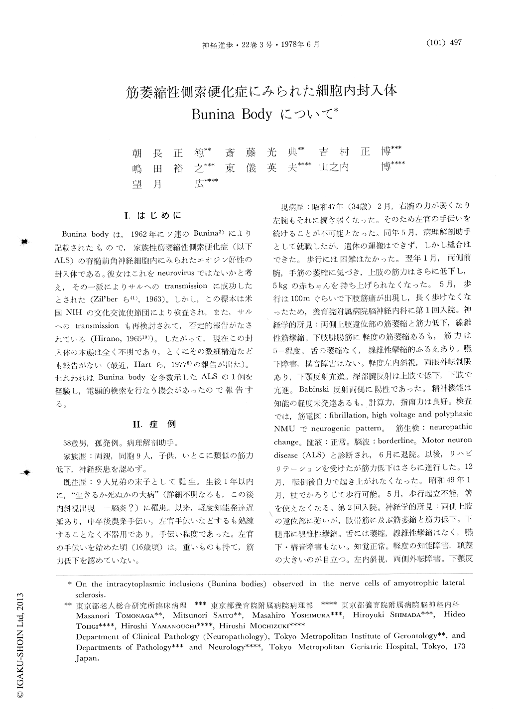
Bunina bodyは,1962年にソ連のBunina3)により記載されたもので,家族性筋萎縮性側索硬化症(以下ALS)の脊髄前角神経細胞内にみられたエオジン好性の封入体である.彼女はこれをneurovirusではないかと考え,その一派によりサルへのtransmissionに成功したとされた(Zil'berら41),1963)。しかし,この標本は米国NIHの文化交流使節団により検査され,また,サルへのtransmissionも再検討されて,否定的報告がなされている(Hirano,196510))。したがって,現在この封入体の本態は全く不明であり,とくにその微細構造なども報告がない(最近,Hartら,19778)の報告が出た)。われわれはBunina bodyを多数示したALSの1例を経験し,電顕的検索を行なう機会があったので報告する。
Abstract
Light and electron microscopic studies were made on the anterior horn cells and brain stem motor neurons in a case of amyotrophic lateral sclerosis. The patient was a man of 38 years who died of respiratory insufficiency. Since he suffered from "encephalitis" (?) at 1 year-old, he showed mental retardation and strabismus. No familial history of any neurological disease. At 34, he found weakness of grip power and wasting of both hands. After 1 year, he developed spastic paraparesis, later showed bulbar sign.
Copyright © 1978, Igaku-Shoin Ltd. All rights reserved.