Japanese
English
- 有料閲覧
PPV購入案内
1,320円 (1,200円+税10%) こちらは、736ページから752ページの文献です。購入には医書.jp本会員登録が必要です。
会員登録完了後に改めて上記「購入サイトへ」を選択してください。
または「購入サイトへ」選択後に「購入ログイン」、「新規会員登録」の順に進んでください。
会員登録について詳しくはをご確認ください。
- Abstract 文献概要
- 1ページ目 Look Inside
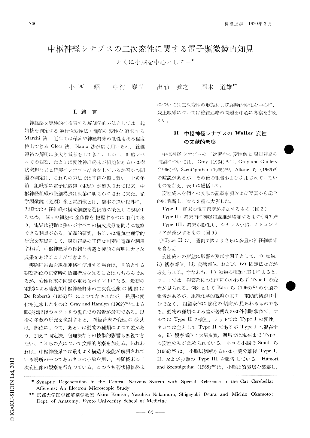
Ⅰ.緒言
神経路を実験的に検索する解剖学的力法としては,起始核を判定する逆行性変性法・髄鞘の変性を追求するMarchi法,近年では軸索や神経終末の変性もある程度検出できるGlees法,Nauta法が広く用いられ,線維連絡の解明に多大な貢献をしてきた。しかし,細胞レベルでの観察,たとえば変性神経終末が細胞体あるいは樹状突起などと確実にシナプス結合をしているか否かの問題の判定は,これらの方法では正確を期し難い。十数年前,組織学に電子顕微鏡(電顕)が導入されて以来,中枢神経組織の微細構造は次第に明らかにされて来た。光学顕微鏡(光顕)像と電顕像とは,倍率の違い以外に,光顕では神経組織の構成細胞を選択的に染色して観察するため,個々の細胞の全体像を把握するのに有利であり,電顕は視野は狭いがすべての構成成分を同時に観察できる利点がある。光顕的研究,あるいは電気生理学的研究を基礎にして,線維連絡の正確な判定に電顕を利用すれば,中枢神経系の複雑な構造と機能の解明に大きな成果をあげることができよう。
A critical assessment was made on the ultrastructure of degenerating synapses described in the literature in many regions and species, referring to our own study on the cat cerebellar afferents after lesions of the olivary nucleus or cerebellar peduncles. Degenerating boutons could be classified into 3 types: type Ⅰ was characterized by increased electron density, type Ⅱ by proliferation of neurofilaments and type Ⅲ by swelling and/or vacuolation. These defferences of the reaction seem to be dependent on the region examined, the site of lesions, the animal species and the mode of specimen preparation.
Copyright © 1970, Igaku-Shoin Ltd. All rights reserved.